
 PV
 TG
 MN
 EM
 EI
 ND

 CT Scan
 PET CT
 Digital X-ray
 Ultrasound
 Amniocentesis

 Fetal Reduction
 Thermalytix Breast Screening
 Cardiology
 Lab Tests
 GAMCA Pre-Medical

A prostate ultrasound is a painless scan that measures the size (volume) of the prostate gland and checks how well the bladder empties. It is usually done through the lower abdomen with a full bladder, and occasionally through the rectum (TRUS) when more detail is needed.
If you are a man over 40 with a weak urine stream, frequent trips to the toilet at night, or a feeling that the bladder never quite empties, your doctor will very likely ask for a prostate ultrasound. Many men then receive a report full of numbers — “prostate volume 48 cc”, “Grade II prostatomegaly”, “post-void residual 120 mL” — with no explanation of what they mean.
This guide explains what a prostate ultrasound shows, how prostate size is measured, what a normal prostate volume is, what post-void residual means, how to read an enlarged prostate ultrasound report, what ultrasound can and cannot tell you about cancer, and when a transrectal scan, MRI or biopsy is the next step. It is general information; your urologist or physician interprets your own results.
Prostate ultrasound at a glance
- What it measures: prostate size (volume), the shape of the gland, how much urine is left after passing urine, and the bladder and kidneys
- Usual method: through the lower abdomen (transabdominal), with a comfortably full bladder
- Time: about 15 to 20 minutes, including the scan after you pass urine
- Preparation: drink water to fill the bladder beforehand; no fasting for a prostate-only scan
- Normal size: roughly 20 to 25 mL in a young adult; it grows slowly with age
- Main limitation: ultrasound cannot reliably detect prostate cancer; a raised PSA usually leads to MRI
- Safety: no radiation, no injection
What this guide covers
- What a prostate ultrasound is
- The prostate and why it enlarges
- When a prostate ultrasound is advised
- Transabdominal ultrasound vs TRUS
- How to prepare
- What happens during the scan
- How prostate size and volume are measured
- Normal prostate size by age
- Prostatomegaly grades on Indian reports
- Post-void residual: what it means
- Other findings on an enlarged prostate ultrasound
- What the scan shows about the bladder and kidneys
- Can ultrasound detect prostate cancer?
- PSA, prostate volume and PSA density
- Ultrasound, TRUS, MRI or biopsy: which test when
- How the results guide treatment
- A sample report, explained
- Prostatitis and prostate abscess on ultrasound
- Prostate ultrasound and male fertility
- Living with an enlarged prostate
- Myths and facts about prostate size
- Urinary symptoms that need urgent attention
- Questions to ask your doctor
- Prostate ultrasound in Delhi
- References
- Frequently asked questions
What a prostate ultrasound is
A prostate ultrasound uses high-frequency sound waves to create live pictures of the prostate gland, the bladder above it and, usually, the kidneys. In most cases the probe is moved over the lower abdomen with a little gel; nothing goes inside the body. The scan has no radiation and needs no injection.
It answers a few practical questions that guide treatment: How big is the prostate? Is part of it bulging up into the bladder? How much urine is left behind after you pass urine? Is the bladder wall or the kidneys showing signs of strain? It is often requested as part of a KUB (kidneys, ureters, bladder) or whole abdomen scan.
The prostate and why it enlarges
The prostate is a gland about the size of a walnut in young men. It sits just below the bladder and wraps around the urethra — the tube that carries urine out of the body. It produces part of the fluid in semen.
From middle age onwards, the prostate gradually enlarges in most men. This non-cancerous growth is called benign prostatic hyperplasia (BPH). Because the prostate surrounds the urethra, an enlarged gland can narrow the outflow of urine, causing symptoms such as:
- A weak or interrupted urine stream
- Difficulty starting to pass urine (hesitancy)
- Needing to pass urine often, including at night (nocturia)
- Urgency, sometimes with leakage
- A feeling that the bladder has not emptied completely
- Dribbling after finishing
These are called lower urinary tract symptoms (LUTS). Not every man with an enlarged prostate has symptoms, and the severity of symptoms does not always match the size of the gland — a moderately enlarged prostate can cause more trouble than a large one, depending on its shape.
When a prostate ultrasound is advised
Doctors commonly request a prostate ultrasound for:
- Lower urinary tract symptoms in men, to measure prostate size and post-void residual
- Recurrent urinary infections, or difficulty passing urine
- An episode of urinary retention (being unable to pass urine)
- Blood in the urine, usually alongside a full kidney and bladder scan
- Planning treatment — some medicines and most procedures depend on prostate size
- Calculating PSA density when the PSA blood test is raised
- Suspected prostate infection or abscess
- Monitoring the effect of treatment over time
European urology guidelines recommend measuring the post-void residual in every man being assessed for LUTS, and imaging the prostate when surgery or a procedure is being considered, or when it helps choose a medicine.
Transabdominal ultrasound vs TRUS
There are two ways to scan the prostate.
| Transabdominal prostate ultrasound | Transrectal ultrasound (TRUS) | |
|---|---|---|
| How | Probe on the lower abdomen, through a full bladder | Slim, lubricated probe placed in the rectum |
| Comfort | Painless | Mild pressure; brief discomfort for some men |
| Volume accuracy | Good for most purposes | More accurate, especially for planning procedures |
| Detail of the gland | Overall size and shape | Finer detail of the gland, seminal vesicles and any abscess |
| Post-void residual and kidneys | Yes, in the same sitting | Not its purpose |
| Typical use | First test for urinary symptoms | Accurate sizing before procedures, prostate abscess, guiding a biopsy |
Guidelines note that transrectal measurement of prostate volume is more accurate than transabdominal measurement. For most men with urinary symptoms, however, the transabdominal scan answers the clinical question. A transrectal scan is used when a precise volume or finer detail is needed. If your doctor has asked for one, our page on the TRUS scan and TRUS prostate biopsy in Delhi explains that procedure.
How to prepare
For a transabdominal prostate ultrasound:
- Fill your bladder. Drink about four to five glasses of water around 45 to 60 minutes before the appointment, and do not pass urine until you are asked. A comfortably full bladder lifts the bowel away and acts as a window to the prostate.
- Fasting is not needed for a prostate-only scan. If a whole abdomen scan is being done at the same time, you may be asked to fast for a few hours beforehand so the gallbladder and pancreas can be seen.
- Bring your prescription, previous ultrasound reports, PSA results and a list of medicines, especially any prostate medicines.
- Wear comfortable clothing that allows easy access to the lower abdomen.
If a transrectal scan has been requested, you will be told separately about any additional preparation.
What happens during the scan
- You lie on your back and a little warm gel is applied to the lower abdomen.
- The prostate is measured in two planes — across (transverse) and from the side (sagittal) — and its shape and texture are assessed.
- The bladder is examined while full: its wall, any stones or pouches (diverticula), and whether the prostate bulges into it.
- The kidneys are checked for swelling (hydronephrosis), stones or other problems.
- You are then asked to empty your bladder completely in the toilet.
- The bladder is scanned again straight away to measure the post-void residual — the urine left behind.
The whole process usually takes about 15 to 20 minutes. Holding a full bladder can be uncomfortable for men with urgency; tell the staff, and the scan can be prioritised.
How prostate size and volume are measured
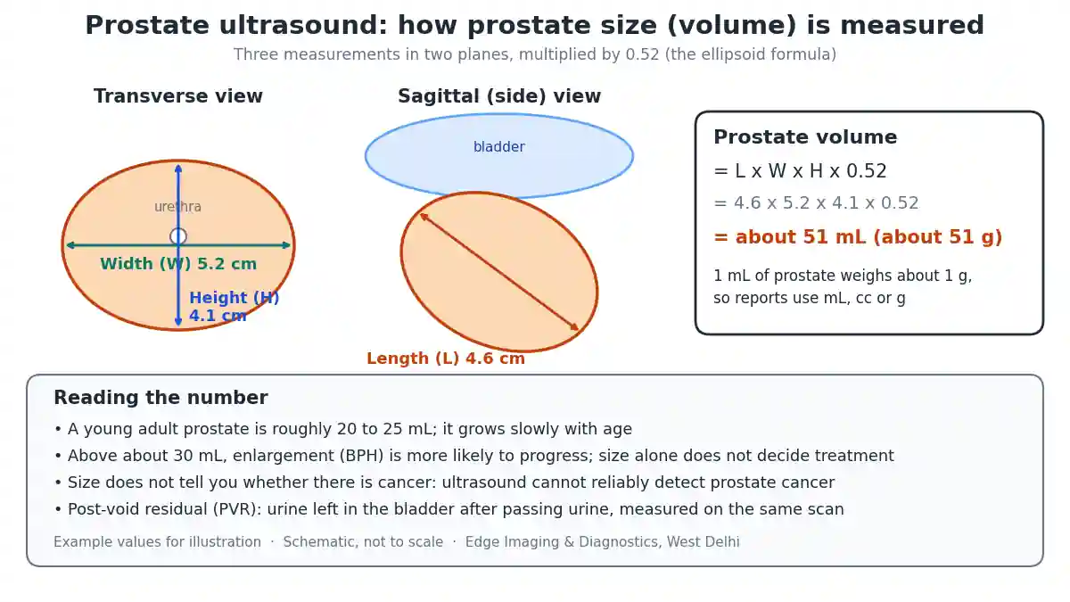
Prostate size is reported as a volume. The sonologist measures three dimensions — length, width and height — and multiplies them by 0.52, a formula that treats the prostate as an ellipsoid (a rounded egg shape).
For example, a prostate measuring 4.6 cm by 5.2 cm by 4.1 cm has a volume of about 4.6 × 5.2 × 4.1 × 0.52, which is roughly 51 mL. Because prostate tissue weighs about 1 gram per millilitre, reports may give the same number in mL, cc or grams — 51 mL, 51 cc and 51 g mean the same thing.
Prostate volume measurement by ultrasound is an estimate. Results can vary by a few millilitres between scans, operators and machines, and the formula is less precise for unusually shaped glands, such as those with a large middle lobe. Small differences between reports are not usually meaningful; large changes over time are.
Normal prostate size by age
There is no single “normal” number for every man, because the prostate grows slowly with age.
| Prostate volume | What it generally suggests |
|---|---|
| About 20 to 25 mL | Typical size in a young adult man |
| About 25 to 30 mL | Commonly seen in middle age; often within the expected range for age |
| Over about 30 mL | Enlarged; BPH is more likely to progress at this size |
| Over about 80 mL | Markedly enlarged; influences the choice of procedure if one is needed |
Two important points. First, size alone does not decide whether treatment is needed — symptoms, post-void residual and complications matter more. Second, a large prostate is not the same as cancer, and a normal-sized prostate does not rule cancer out.
Prostatomegaly grades on Indian reports
Many reports in India describe an enlarged prostate as Grade I, Grade II or Grade III prostatomegaly, sometimes with a Grade IV for very large glands. These grades are a convenient shorthand, but the volume ranges used for each grade differ from one centre and textbook to another. That means “Grade II” at one centre may not be exactly the same size as “Grade II” at another.
The actual volume in mL or grams is far more useful than the grade, especially when comparing reports from different centres over the years. If your report gives only a grade, ask whether the volume can be stated as well.
Post-void residual: what it means
The post-void residual (PVR) is the amount of urine left in the bladder immediately after you have passed urine as completely as you can. It is measured by scanning the bladder again within a few minutes of emptying.
| Post-void residual | How it is generally viewed |
|---|---|
| Very small (a few tens of mL) | Good bladder emptying |
| Up to about 100 to 150 mL | Often within the range regarded as acceptable, depending on age and symptoms |
| Over about 200 mL | Generally regarded as significant incomplete emptying |
| Very large volumes | Risk of retention, infection and kidney strain; needs prompt review |
A few practical cautions:
- A single raised PVR can be misleading — if you were anxious, had over-filled the bladder or could not empty properly in an unfamiliar toilet, it may be repeated.
- A raised PVR does not prove the prostate is blocking the outflow. A weak bladder muscle can also leave urine behind.
- European guidelines note that no single PVR threshold has been established for deciding treatment, but serial measurements can help identify men at risk of retention.
Other findings on an enlarged prostate ultrasound
An enlarged prostate ultrasound report often mentions some of the following.
- Median lobe or intravesical prostatic protrusion (IPP): part of the prostate bulging up into the base of the bladder, measured in millimetres. Guidelines grade it as under 5 mm, 5 to 10 mm, and over 10 mm; a larger protrusion correlates with outflow obstruction and can affect the choice of procedure.
- Prostatic calcifications: small bright spots of calcium. Very common with age and usually harmless.
- Small cysts: usually incidental and harmless.
- Echotexture: descriptions such as “heterogeneous” are common in enlarged glands and are not in themselves a sign of cancer.
- Abscess: a fluid collection in the prostate, usually with fever and pain; a transrectal scan shows it best.
What the scan shows about the bladder and kidneys
Because urinary outflow problems affect the whole urinary tract, the scan also looks at:
- The bladder wall: long-standing obstruction can make the wall thick and ridged (trabeculated). Wall thickness measurements are not standardised, so guidelines do not recommend relying on them alone.
- Bladder diverticula: pouches that can form in a bladder working against obstruction.
- Bladder stones: which can form when urine stagnates.
- The kidneys: swelling (hydronephrosis) suggests urine is backing up and needs prompt attention. Kidney stones are also looked for — see our guide to which scan is best for kidney stone pain.
Can ultrasound detect prostate cancer?
This is one of the most common misunderstandings. A standard prostate ultrasound cannot reliably detect or rule out prostate cancer. European prostate cancer guidelines state plainly that standard TRUS is not reliable for detecting prostate cancer, and most cancers look the same as normal tissue on ordinary ultrasound.
So a “normal” prostate ultrasound does not mean you do not have cancer, and an enlarged prostate does not mean you do. Prostate cancer is assessed with the PSA blood test, a rectal examination by your doctor, MRI of the prostate, and, when needed, a biopsy.
PSA, prostate volume and PSA density
PSA (prostate-specific antigen) is a protein made by the prostate. It rises with prostate cancer, but also with BPH, infection, recent ejaculation, a recent catheter and simply with a larger gland. A benign prostate of around 35 mL typically produces a PSA in the region of 1.5 ng/mL, so a big gland can produce a higher PSA without cancer.
This is where the ultrasound volume becomes useful. Dividing the PSA by the prostate volume gives the PSA density. A PSA that is high for the size of the gland is more suspicious than the same PSA in a very large gland. Guidelines note that PSA density remains predictive when combined with MRI findings. Your urologist interprets it together with your age, family history, examination and MRI.
Ultrasound, TRUS, MRI or biopsy: which test when
| Situation | Usual test |
|---|---|
| Urinary symptoms, suspected enlarged prostate | Transabdominal prostate ultrasound with post-void residual, plus kidney and bladder scan |
| Precise volume before a procedure | Transrectal ultrasound (TRUS) or MRI |
| Raised PSA or abnormal rectal examination | MRI of the prostate, usually before any biopsy — see MRI pelvis |
| Suspicious area on MRI | Targeted biopsy, such as MRI-fusion prostate biopsy |
| Biopsy advised by the urologist | Prostate biopsy, guided by TRUS |
| Fever, pain and suspected prostate abscess | Transrectal ultrasound and urgent clinical review |
European guidelines recommend that men with a PSA between 3 and 20 ng/mL and no symptoms should have their need for biopsy assessed using tools such as MRI of the prostate, risk calculators or additional biomarker tests, rather than going straight to a biopsy.
How the results guide treatment
The ultrasound findings feed into treatment decisions made by your doctor:
- Watchful waiting and lifestyle changes for mild symptoms with good bladder emptying — reducing evening fluids, caffeine and alcohol, and bladder training
- Medicines that relax the prostate (alpha-blockers) for bothersome symptoms
- Medicines that shrink the prostate (5-alpha-reductase inhibitors), which work mainly in larger glands — this is one reason prostate size is measured before starting them
- Procedures or surgery when symptoms are severe, medicines fail, or there are complications such as retention, recurrent infection, stones or kidney strain; prostate size and the presence of a median lobe help decide which procedure is suitable
Your doctor will usually also use a symptom questionnaire, such as the International Prostate Symptom Score, a urine test, a kidney function blood test and sometimes a urine flow test. The ultrasound is one piece of this picture: it adds objective numbers, but treatment is chosen on how much the symptoms bother you and whether there are complications.
Repeat scans over time can show whether the prostate is growing and whether bladder emptying is improving on treatment.
A sample report, explained
Here is an illustrative example of an enlarged prostate ultrasound report and what each line means.
| Report line | What it means |
|---|---|
| Prostate enlarged, measures 4.6 × 5.2 × 4.1 cm, volume about 51 cc | The prostate is about twice the size of a young adult’s gland |
| Heterogeneous echotexture with a few calcific foci | Common in enlarged glands; calcium spots are usually harmless |
| Intravesical prostatic protrusion 8 mm | A moderate bulge into the bladder base; relevant to outflow obstruction |
| Bladder wall mildly thickened and trabeculated | Signs that the bladder has been working against resistance |
| Pre-void volume 380 mL; post-void residual 140 mL | The bladder is not emptying completely; your doctor will consider symptoms and repeat measurements |
| Both kidneys normal, no hydronephrosis | No sign of urine backing up into the kidneys |
This example is for illustration only. Your own report should always be interpreted by your doctor, together with your symptoms and other tests.
Prostatitis and prostate abscess on ultrasound
Prostatitis means inflammation of the prostate, usually from infection. Acute bacterial prostatitis causes fever, chills, pain between the legs or in the lower back, burning when passing urine and sometimes difficulty passing urine. It is diagnosed mainly by the doctor’s examination and urine tests, and ultrasound is often normal or shows only a slightly swollen gland.
Ultrasound becomes important when the infection does not settle with antibiotics as expected. Then the concern is a prostate abscess — a collection of pus within the gland. A transrectal scan shows an abscess best, and it may need drainage. Chronic prostatitis and chronic pelvic pain are usually diagnosed clinically; ultrasound mainly helps exclude other causes and measure bladder emptying.
Prostate ultrasound and male fertility
Ultrasound of the prostate region is occasionally used in the assessment of male infertility. In men with a very low semen volume and no sperm, a transrectal scan can look for blockage of the ejaculatory ducts, enlarged seminal vesicles or cysts near the midline of the prostate, which may be treatable. This is a specialised use requested by a urologist or andrologist after semen analysis, and is different from the routine scan done for urinary symptoms.
Living with an enlarged prostate
For many men with an enlarged prostate and mild symptoms, simple changes make a real difference, and your doctor may suggest trying them before or alongside medicines:
- Reducing fluids in the two hours before bed, while drinking enough during the day
- Cutting down on caffeine and alcohol, which irritate the bladder and increase urine production
- Double voiding — waiting a moment after passing urine and then trying again
- Bladder training to gradually lengthen the time between toilet visits
- Reviewing medicines that can worsen symptoms, such as some cold remedies and certain antihistamines
- Avoiding constipation, which can press on the bladder
- Staying active and keeping a healthy weight
These measures do not shrink the prostate, but they can ease symptoms and improve quality of life. Repeat ultrasound scans help confirm that bladder emptying is stable over time.
Myths and facts about prostate size
| Myth | Fact |
|---|---|
| A bigger prostate always causes worse symptoms | Symptoms depend on shape and bladder function too; a moderately enlarged gland with a median lobe can cause more trouble than a larger one |
| An enlarged prostate means cancer | Enlargement with age is usually benign prostatic hyperplasia, which is not cancer |
| A normal ultrasound rules out prostate cancer | Standard ultrasound cannot reliably detect prostate cancer; PSA, examination and MRI are used |
| The grade on the report is all that matters | The volume in mL and the post-void residual are more useful, and grades vary between centres |
| Some urine left in the bladder is always dangerous | Small residual volumes are common; large or rising volumes need attention |
| Once enlarged, nothing can be done | Lifestyle measures, medicines and procedures can all relieve symptoms |
Urinary symptoms that need urgent attention
Seek medical help promptly, without waiting for a routine scan, if you have:
- Inability to pass urine, especially with a painful, swollen lower abdomen (acute urinary retention)
- Fever, chills or pain in the lower back or between the legs with urinary symptoms
- Visible blood in the urine
- Passing very little urine, swelling of the legs or feeling unwell with known retention
Questions to ask your doctor
- What is my prostate volume, and how has it changed since my last scan?
- How much urine is left after I pass urine, and is that a concern?
- Is there a median lobe or protrusion into the bladder?
- Are my kidneys and bladder affected?
- Does my PSA need further assessment, such as an MRI?
- Which treatment do you recommend, and how will we monitor it?
- When should I have a repeat scan?
Prostate ultrasound in Delhi
Edge Imaging & Diagnostics is a NABH-accredited diagnostic group in West Delhi. We perform prostate ultrasound with post-void residual measurement, KUB and whole abdomen ultrasound, transrectal ultrasound (TRUS), MRI of the prostate and pelvis, and image-guided prostate biopsy, including TRUS biopsy. Reports state the prostate volume in mL, the post-void residual and the kidney findings, and are prepared by our consultant radiologists.
Send a photograph of your prescription on WhatsApp and we will tell you which scan has been asked for, how to prepare, and today’s actual pricing.
💬 Send your prescription on WhatsApp for preparation and today’s pricing | Call 9555999960
Related reading: TRUS scan and prostate biopsy centre · Prostate biopsy in Delhi · MRI-fusion prostate biopsy · MRI pelvis · Whole abdomen ultrasound · Testicular pain or swelling: which scan
References
- Management of Non-neurogenic Male LUTS: Diagnostic Evaluation — European Association of Urology (EAU) Guidelines.
- Prostate Cancer: Diagnostic Evaluation — European Association of Urology (EAU) Guidelines.
- Benign Prostatic Hyperplasia — StatPearls, NCBI Bookshelf (US National Library of Medicine).
- Transrectal Ultrasound in Prostate Cancer: Current Utilization, Integration with mpMRI, HIFU and Other Emerging Applications — Panzone J, et al. Cancer Management and Research, 2022.
Frequently asked questions
What is a prostate ultrasound?
A prostate ultrasound is a painless scan that measures the size of the prostate gland and checks how well the bladder empties. It is usually done through the lower abdomen with a full bladder, and occasionally through the rectum (TRUS) when more detail is needed.
What is the normal prostate size?
In a young adult man the prostate is roughly 20 to 25 mL (about 20 to 25 grams). It grows slowly with age, and a prostate above about 30 mL is generally regarded as enlarged. Size alone does not decide whether treatment is needed.
How is prostate volume calculated on ultrasound?
The length, width and height of the prostate are measured and multiplied by 0.52, the ellipsoid formula. For example, 4.6 by 5.2 by 4.1 cm gives about 51 mL. Because prostate tissue weighs about 1 gram per mL, the result may be reported in mL, cc or grams.
What does post-void residual mean?
Post-void residual is the amount of urine left in the bladder immediately after you pass urine, measured by scanning the bladder again. Small amounts are normal; volumes over about 200 mL are generally regarded as significant incomplete emptying, though a single reading may need repeating.
How should I prepare for a prostate ultrasound?
Drink about four to five glasses of water 45 to 60 minutes before the scan and do not pass urine until asked. Fasting is not needed for a prostate-only scan, but may be needed if a whole abdomen scan is done at the same time.
Is a prostate ultrasound painful?
No. The transabdominal scan is painless; the only discomfort is holding a full bladder. A transrectal scan causes mild pressure and brief discomfort for some men, but is generally well tolerated.
Can a prostate ultrasound detect prostate cancer?
Not reliably. Most prostate cancers look like normal tissue on standard ultrasound, and guidelines state that standard TRUS is not reliable for detecting prostate cancer. A raised PSA is usually followed by MRI of the prostate and, if needed, a biopsy.
What is the difference between abdominal prostate ultrasound and TRUS?
An abdominal scan places the probe on the lower abdomen through a full bladder and also measures post-void residual and checks the kidneys. TRUS uses a slim probe in the rectum and gives a more accurate volume and finer detail, used before procedures, for abscesses or to guide a biopsy.
What does Grade II prostatomegaly mean?
It means the prostate is moderately enlarged. However, the volume ranges used for each grade differ between centres, so the actual volume in mL or grams is more useful than the grade, especially when comparing reports over time.
What is intravesical prostatic protrusion?
It is part of the prostate bulging upwards into the base of the bladder, measured in millimetres on ultrasound. A protrusion of more than 10 mm correlates with obstruction of urine outflow and can influence the choice of treatment.
Does a big prostate mean cancer?
No. Prostate enlargement with age is usually benign prostatic hyperplasia, which is not cancer. A large prostate does not mean cancer, and a normal-sized prostate does not rule cancer out.
What is PSA density?
PSA density is the PSA blood level divided by the prostate volume measured on ultrasound or MRI. A PSA that is high for the size of the gland is more suspicious than the same PSA in a very large gland, and doctors interpret it together with MRI and other factors.
Why do I need to empty my bladder during the scan?
So that the post-void residual can be measured. The bladder is scanned while full to see the prostate and bladder clearly, then again immediately after you pass urine to see how much is left behind.
Do I need to fast before a prostate ultrasound?
Not for a prostate-only scan. If your prescription includes a whole abdomen ultrasound, you may be asked to fast for a few hours beforehand so the gallbladder and pancreas can be seen properly.
How often should a prostate ultrasound be repeated?
There is no fixed interval. Your doctor may repeat it to check the response to treatment, to monitor post-void residual, or if symptoms change. Comparing the volume in mL between reports is the most useful way to track change.
What symptoms need urgent attention?
Seek urgent help for inability to pass urine with a painful swollen lower abdomen, fever or chills with urinary symptoms, visible blood in the urine, or feeling unwell while passing very little urine.
Where can I get a prostate ultrasound in Delhi?
Edge Imaging & Diagnostics in West Delhi performs prostate ultrasound with post-void residual, KUB and whole abdomen ultrasound, TRUS, prostate MRI and image-guided prostate biopsy. Send your prescription on WhatsApp to 9555999960 for preparation and today’s pricing.
The bottom line
A prostate ultrasound is a quick, painless scan that measures prostate size and how well your bladder empties. The volume in mL, the post-void residual and the state of the bladder and kidneys are the numbers that matter most for treatment.
It cannot reliably detect prostate cancer: a raised PSA leads to MRI and, if needed, a biopsy. Bring your previous reports so changes can be compared, and let your doctor put the numbers in context.
Written by the clinical content team at Edge Imaging & Diagnostics, West Delhi — a NABH-accredited diagnostic centre. Medically reviewed by Dr. Nishant Thapar, Consultant Radiologist. Last reviewed: October 2026.
Medical disclaimer: this article is general information about prostate ultrasound. It is not medical advice or a diagnosis. Your doctor interprets your results together with your symptoms and other tests.
Size ranges, thresholds and examples quoted here are approximate and vary between guidelines, centres and individuals. The sample report is illustrative and not a real patient’s report.