
 PV
 TG
 MN
 EM
 EI
 ND

 CT Scan
 PET CT
 Digital X-ray
 Ultrasound
 Amniocentesis

 Fetal Reduction
 Thermalytix Breast Screening
 Cardiology
 Lab Tests
 GAMCA Pre-Medical

Testicular pain or swelling has many causes, from harmless fluid collections to emergencies. Sudden, severe testicular pain may be testicular torsion and needs hospital care immediately. For most other pain, swelling or a lump, the right first test is a scrotal ultrasound with Doppler.
Pain or swelling in the testicles is something many men feel embarrassed to talk about, and so they wait. Sometimes waiting is harmless. Sometimes it costs a testicle. This guide explains, in plain language, the common causes of testicular pain, a swollen testicle and a testicular lump, how to tell when it is an emergency, what the scan shows, and what usually happens next.
It is general information, not a diagnosis. If you are worried, see a doctor — and if the pain is sudden and severe, go to hospital now rather than reading further.
Testicular pain at a glance
- Sudden, severe pain: treat as testicular torsion until proven otherwise — emergency surgery within about 6 hours saves the testicle in 90 to 100% of cases
- Pain building over hours to days, with fever or burning urine: often epididymitis or orchitis — same-day medical review
- A painless lump or a hard, heavy testicle: needs a prompt scrotal ultrasound; testicular cancer must be excluded
- A soft swelling or “bag of worms”: often a hydrocele, varicocele or cyst — usually not urgent, but should be scanned
- The main test: scrotal ultrasound with colour Doppler — painless, quick and radiation-free
What this guide covers
- Is it an emergency? Check this first
- The testicle and scrotum in brief
- Causes of testicular pain and swelling at a glance
- Testicular torsion
- Torsion of the appendix testis
- Epididymitis and orchitis
- Injury to the testicle
- Hydrocele
- Varicocele
- Epididymal cysts and spermatocele
- Inguinal hernia
- A testicular lump and testicular cancer
- Pain felt in the testicle from elsewhere
- Long-standing testicular pain
- A rare emergency: severe infection of the scrotal skin
- Undescended and retractile testicles
- Testicular microlithiasis explained
- Testicular pain after vasectomy or hernia surgery
- What if the scan is normal?
- Which scan do you need?
- What happens during a scrotal ultrasound
- What the scan can show
- Other tests your doctor may order
- Testicular pain in boys and teenagers
- How to examine your testicles
- Questions to ask your doctor
- Scrotal ultrasound in Delhi
- References
- Frequently asked questions
Is it an emergency? Check this first
Go to a hospital emergency department immediately — do not wait for a routine scan — if you have:
- Sudden, severe pain in one testicle
- Testicular pain with nausea, vomiting or pain in the lower abdomen
- Testicular pain that has lasted more than an hour, or continues when you rest
- A testicle that sits higher than usual or lies at an odd angle
- Severe pain or rapid swelling after an injury
- Painful, red, rapidly spreading swelling of the scrotum with fever, especially in diabetics
These can be signs of testicular torsion, a ruptured testicle or a serious infection. In torsion, time is everything. If a specialist strongly suspects torsion, surgery should not be delayed to get a scan. Ask someone else to drive you, or call an ambulance.
The testicle and scrotum in brief
Each testicle (testis) sits in the scrotum, hanging from the spermatic cord, which carries its blood vessels and the vas deferens. Behind each testicle lies the epididymis, a coiled tube where sperm mature. The testicle is surrounded by a thin double layer of tissue that normally contains a tiny amount of fluid. Problems in any of these structures — the testicle, the epididymis, the cord, the veins or the surrounding fluid — can cause pain or swelling, and the scrotal ultrasound is designed to look at each of them.
Causes of testicular pain and swelling at a glance
| Cause | Typical pattern | Urgency |
|---|---|---|
| Testicular torsion | Sudden severe pain, often with vomiting; mostly teenagers and young men | Emergency |
| Torsion of the appendix testis | Pain at the top of the testicle, usually in boys; more gradual | Urgent assessment |
| Epididymitis or orchitis | Pain and swelling building over a day or more, sometimes fever or urinary symptoms | Same-day review |
| Injury | Pain and swelling after a blow | Urgent if severe |
| Hydrocele | Painless, soft, smooth swelling | Routine scan |
| Varicocele | Dull ache, “bag of worms” above the testicle, usually left side | Routine scan |
| Epididymal cyst or spermatocele | Small smooth lump separate from the testicle | Routine scan |
| Inguinal hernia | Groin bulge extending into the scrotum, larger on coughing | Urgent if painful and cannot be pushed back |
| Testicular cancer | Usually a painless lump or a hard, heavy testicle | Prompt scan, within days |
| Referred pain | Pain felt in the testicle from a kidney stone, hernia or back problem | Depends on cause |
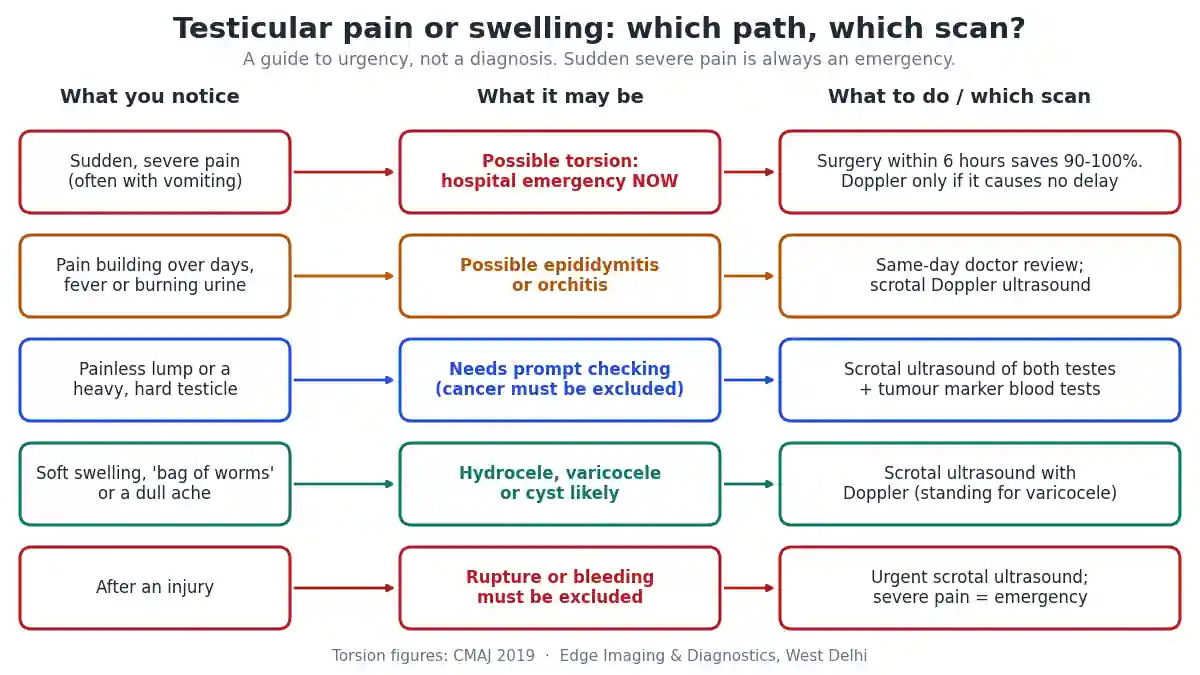
Testicular torsion
Testicular torsion happens when the testicle rotates and twists its spermatic cord, cutting off its own blood supply. It mainly affects pubertal boys and young men, occurring in about 1 in 4,000 males under 25, but it can happen at any age.
Typical symptoms: sudden, severe pain in one testicle, often waking a young man from sleep or starting during activity; nausea and vomiting; pain in the lower abdomen; a swollen, very tender testicle that may sit higher than the other. Some men have had brief, self-resolving episodes of similar pain before.
Why speed matters: the testicle can survive only a few hours without blood. When surgery is performed within about 6 hours of the pain starting, the testicle is saved in 90 to 100% of cases. At 12 to 24 hours, salvage falls to around 10%.
The role of the scan: colour Doppler ultrasound is the imaging test of choice. It shows reduced or absent blood flow in the twisted testicle, and sometimes a “whirlpool sign” of the twisted cord. Its sensitivity is high but not perfect, and a testicle that has twisted and untwisted may look nearly normal. That is why guidelines say that when torsion is strongly suspected, surgery should not be delayed to wait for an ultrasound.
Treatment: an operation to untwist the testicle and fix it in place, usually fixing the other testicle too, because the anatomical tendency to twist is often present on both sides. If the testicle cannot be saved, it is removed.
Torsion of the appendix testis
The appendix testis is a tiny, harmless nub of tissue on the top of the testicle. In children and young teenagers it can twist, causing pain that tends to come on more gradually than true torsion and is focused on the upper part of the testicle. Sometimes a small blue dot is visible through the skin. A Doppler ultrasound helps distinguish it from testicular torsion by showing normal blood flow to the testicle. It usually settles with rest and painkillers, but because it can mimic torsion, it always needs medical assessment.
Epididymitis and orchitis
Epididymitis is inflammation of the epididymis, usually from infection; when the testicle is involved too, it is called epididymo-orchitis. It typically causes pain and swelling that build up over hours to days rather than seconds, tenderness at the back of the testicle, and sometimes fever, burning when passing urine or a discharge.
- In sexually active younger men, sexually transmitted infections such as chlamydia and gonorrhoea are the leading causes.
- In older men, bacteria from the urinary tract, such as E. coli, are more common, often linked with prostate enlargement or a catheter.
- Mumps orchitis can affect the testicle a few days after the swelling of the salivary glands in mumps.
On a Doppler ultrasound, the inflamed epididymis or testicle is swollen and shows increased blood flow — the opposite of torsion. Treatment is with antibiotics chosen for the likely organism, rest, scrotal support and painkillers. Untreated or late-treated infection can lead to an abscess, which a repeat scan can detect, so return promptly if you are not improving.
Injury to the testicle
A blow to the scrotum, from sport, a fall or an accident, can cause bruising, a collection of blood (haematocele), or a rupture of the tough outer covering of the testicle. Severe pain, rapid swelling or a testicle that feels abnormal after an injury needs urgent assessment. A scrotal ultrasound shows whether the testicle’s outline is intact and how much bleeding there is. A ruptured testicle usually needs prompt surgery to save it.
Hydrocele
A hydrocele is a collection of fluid around the testicle. It causes a smooth, soft, usually painless swelling that can become large and heavy. In adults it may follow infection or injury, or have no obvious cause; in babies it is common and often settles on its own.
Ultrasound confirms the fluid and, importantly, shows the testicle inside, which cannot always be felt through a large hydrocele. This matters because, occasionally, a hydrocele forms around a testicle with another problem. Treatment is needed only if the hydrocele is large, uncomfortable or troublesome, usually with a minor operation.
Varicocele
A varicocele is a group of swollen veins above the testicle, rather like varicose veins in the legs. It is common, usually on the left side, and often feels like a “bag of worms” when standing. It may cause a dull, dragging ache, worse at the end of the day or after standing for long periods.
On a Doppler ultrasound, typically performed lying down and then standing or while straining, the veins are wider than usual — often more than about 3 mm — with blood flowing backwards on straining. Varicoceles are linked with reduced fertility in some men and with a smaller testicle on the affected side in adolescents. Many need no treatment; treatment is considered for pain, infertility, or a shrinking testicle in a teenager. A new varicocele that appears suddenly in an older man, especially on the right side or one that does not empty when lying down, needs further assessment of the abdomen.
Epididymal cysts and spermatocele
An epididymal cyst or spermatocele is a fluid-filled sac in the epididymis, felt as a small, smooth lump that is separate from the testicle, usually above or behind it. They are very common, harmless and usually painless. Ultrasound confirms the diagnosis in seconds, which is often the main reassurance a man needs. Treatment is only required if a cyst becomes large or uncomfortable.
Inguinal hernia
An inguinal hernia is a bulge of abdominal contents through a weak point in the groin, which can extend down into the scrotum. It often gets bigger on coughing or straining and may be pushed back when lying down. A hernia that becomes painful, firm and cannot be pushed back, especially with vomiting, may be strangulated and is an emergency. Ultrasound can show bowel or fat in the scrotum and help distinguish a hernia from other swellings.
A testicular lump and testicular cancer
Most lumps in the scrotum are not cancer — cysts, hydroceles and varicoceles are far more common. But a lump within the testicle itself must always be checked, because testicular cancer is one of the commonest cancers in young men and is highly curable, especially when found early.
Typical features of testicular cancer:
- A painless lump or swelling in one testicle
- A testicle that has become larger, firmer or heavier
- An ache in the testicle, scrotum or lower abdomen
- Rarely, back pain, breathlessness or breast swelling from spread or hormone effects
Pain alone is not usually a sign of testicular cancer. The first test for any testicular lump is a scrotal ultrasound of both testicles, which European urology guidelines recommend in every man with suspected testicular cancer, using a high-frequency probe. Ultrasound distinguishes a solid lump inside the testicle from a cyst or fluid outside it. If a solid testicular lump is found, blood tests for tumour markers — AFP, beta-hCG and LDH — are taken, and a urologist plans the next step, which is usually removal of the testicle through the groin for diagnosis and treatment. A needle biopsy of a suspected testicular cancer is generally avoided.
Very small incidental spots found in the testicle on ultrasound are usually benign, and are followed up as your doctor advises.
Pain felt in the testicle from elsewhere
Sometimes the testicle hurts even though nothing is wrong with it. The nerves supplying the testicle share pathways with other areas, so pain can be referred from:
- A kidney or ureteric stone — pain often starts in the side or back and travels to the groin and testicle; see our guide to which scan is best for kidney stone pain
- An inguinal hernia
- Back problems irritating the nerves
- Rarely, an abdominal aortic aneurysm in older men
When the scrotal ultrasound is normal and the pain continues, an abdominal or kidney ultrasound may be the next step.
Long-standing testicular pain
Pain in one or both testicles that lasts for three months or more is called chronic scrotal pain or orchialgia. Causes include a varicocele, a cyst, previous infection or injury, nerve irritation, pain after a vasectomy or hernia repair, and referred pain. In many men no single cause is found. A scrotal ultrasound with Doppler is the starting point to look for a treatable cause, and further management may involve a urologist and a pain specialist. Chronic pain is real and treatable; it should not be dismissed.
A rare emergency: severe infection of the scrotal skin
Very rarely, an infection of the skin and tissues of the scrotum and groin spreads rapidly. This condition, known as Fournier gangrene, is more likely in men with diabetes, a weakened immune system, heavy alcohol use or recent surgery in the area. Warning signs are intense pain and swelling that spread quickly, red, purple or dark skin, a foul smell, crackling under the skin, fever and feeling very unwell. It is a surgical emergency that needs hospital treatment immediately; do not wait for a scan.
Undescended and retractile testicles
In some boys a testicle does not descend fully into the scrotum before birth. An undescended testicle is usually found at birth or during childhood checks and is generally corrected with surgery in early childhood, because it carries a higher risk of reduced fertility and of testicular cancer later in life. A retractile testicle moves up into the groin with cold or touch but can be brought down; it is usually normal but is monitored. Men who had an undescended testicle should be especially careful with monthly self-examination. Ultrasound can help locate a testicle in the groin, although a specialist examination is the key assessment.
Testicular microlithiasis explained
Testicular microlithiasis means multiple tiny bright spots of calcium scattered within the testicle on ultrasound. It is usually found by chance and causes no symptoms. On its own it is generally regarded as a benign finding. Your doctor may advise monthly self-examination, and further follow-up depends on whether there are other risk factors, such as a previous undescended testicle, a small testicle or a personal or family history of testicular cancer. Do not panic if the word appears on your report; ask your doctor what it means in your case.
Testicular pain after vasectomy or hernia surgery
Some discomfort after a vasectomy or an inguinal hernia repair is normal for a few days to weeks. A small number of men develop longer-lasting pain, sometimes from congestion of the epididymis, a small sperm granuloma, scar tissue or irritation of a nerve. A scrotal ultrasound with Doppler helps look for a treatable cause, such as a collection, a cyst or swelling of the epididymis, and the operating surgeon or a urologist plans management.
What if the scan is normal?
A normal scrotal ultrasound is reassuring: it effectively rules out a hydrocele, a solid lump in the testicle, an abscess and a significant varicocele, and shows normal blood flow. If pain continues, the cause may lie elsewhere — a kidney stone, a hernia, the back or a nerve — and your doctor may arrange a kidney or abdominal scan, or a referral to a urologist. Continuing or worsening symptoms always deserve review, even after a normal scan.
Which scan do you need?
| Situation | Usual test |
|---|---|
| Sudden severe pain, possible torsion | Emergency surgical review; Doppler ultrasound only if it does not delay surgery |
| Pain and swelling over days, possible infection | Scrotal ultrasound with colour Doppler, plus urine tests |
| Testicular lump or hard testicle | Scrotal ultrasound of both testicles, then tumour markers if a solid lump is found |
| Soft swelling, possible hydrocele or cyst | Scrotal ultrasound |
| Ache with suspected varicocele | Scrotal Doppler ultrasound, lying and standing or straining |
| Groin bulge into the scrotum | Ultrasound of the groin and scrotum |
| Normal scrotal scan with continuing pain | Kidney or abdominal ultrasound to look for referred causes |
| Ultrasound findings unclear | MRI of the scrotum or pelvis in selected cases |
For almost every non-emergency situation, a scrotal ultrasound (testis ultrasound) with Doppler is the right first test. CT scans are not used to look at the testicles themselves; they have a role mainly in staging testicular cancer.
What happens during a scrotal ultrasound
- You lie on your back on the couch. The penis is moved up onto the abdomen and covered, and a towel is placed under the scrotum to support it. Privacy is maintained throughout.
- Warm gel is applied and a high-frequency probe is moved gently over each side of the scrotum.
- Both testicles are always examined and compared — size, texture and any lumps — along with the epididymis and any fluid.
- Colour Doppler shows blood flow in each testicle and in the veins above them.
- For a suspected varicocele, you may be asked to strain (as if passing a stool) or to stand.
The scan usually takes 15 to 20 minutes and needs no preparation. It is painless, although pressing over a very tender area may be uncomfortable; the sonologist will be as gentle as possible. There is no radiation.
What the scan can show
- Blood flow: absent or reduced in torsion; increased in infection
- Testicle size and texture: including any solid lump within the testicle
- The epididymis: swelling from infection, or cysts
- Fluid: a hydrocele, or blood after injury
- Veins: width and backflow in a varicocele
- Abscess: a pus collection complicating infection
- Hernia contents: bowel or fat in the scrotum
- Small bright spots: tiny calcifications (microlithiasis), usually followed as advised
The report should describe both testicles, give their sizes, and state the Doppler findings clearly, so that your doctor can decide the next step.
Other tests your doctor may order
- Urine tests for infection, and swabs or urine tests for sexually transmitted infections
- Blood tests, including inflammatory markers when infection is suspected
- Tumour markers (AFP, beta-hCG, LDH) when a solid testicular lump is found
- Semen analysis when a varicocele and fertility concerns go together
- MRI of the scrotum or pelvis occasionally, when ultrasound is inconclusive — see our MRI pelvis guide
Testicular pain in boys and teenagers
In boys and teenagers, sudden testicular pain should always be treated as possible torsion. Teenage boys are often embarrassed and may describe only “stomach pain” or vomiting, so parents should ask directly about the testicles when a boy has sudden lower abdominal pain. Torsion of the appendix testis and epididymitis also occur in this age group. In younger children, a painless scrotal swelling is often a hydrocele or hernia. Any doubt means a prompt medical review.
How to examine your testicles
Getting to know what is normal for you makes it easier to notice a change early. Once a month, ideally after a warm bath or shower when the scrotum is relaxed:
- Hold the scrotum in the palm of your hand.
- Gently roll each testicle between your thumb and fingers, feeling for lumps, hard areas or changes in size or shape.
- Find the epididymis — the soft, slightly lumpy tube at the back of each testicle. It is normal, so get to know how it feels.
- It is normal for one testicle to be slightly larger or to hang lower than the other.
If you notice a new lump, swelling, hardness or a change in size, see a doctor and ask for a scrotal ultrasound. Most changes turn out to be harmless, but checking early is what makes testicular cancer so curable.
Questions to ask your doctor
- Could this be torsion, and does it need emergency surgical review?
- Is the problem inside the testicle or outside it?
- What does the Doppler blood flow show?
- Do I need antibiotics, and should my partner be tested?
- Is a solid lump present, and do I need tumour marker tests?
- Is any treatment needed for a hydrocele, varicocele or cyst, or can it be watched?
- When should the scan be repeated?
Scrotal ultrasound in Delhi
Edge Imaging & Diagnostics is a NABH-accredited diagnostic group in West Delhi. We perform scrotal and testicular ultrasound with colour Doppler using high-frequency probes, with both testicles assessed and compared, as well as kidney and whole abdomen ultrasound and MRI for further assessment. Reports are prepared by our consultant radiologists and describe the size, structure and blood flow of both testicles clearly for your doctor. Our detailed page on scrotal ultrasound in Delhi explains the test itself.
If your pain is sudden and severe, do not book a scan — go to the nearest hospital emergency department now.
For non-emergency pain, swelling or a lump, send a photograph of your prescription on WhatsApp and we will tell you which scan has been asked for, the earliest slot, and today’s actual pricing.
💬 Send your prescription on WhatsApp for the earliest slot and today’s pricing | Call 9555999960
Related reading: Scrotal ultrasound in Delhi · Prostate ultrasound · Penile Doppler test · MRI pelvis
References
- Five things to know about testicular torsion — Keays M, Rosenberg H. CMAJ, 2019.
- Epididymitis — StatPearls, NCBI Bookshelf (US National Library of Medicine).
- Testicular Cancer: Diagnostic Evaluation — European Association of Urology (EAU) Guidelines.
- Testicle pain — NHS.
Frequently asked questions
What causes testicular pain?
Common causes include testicular torsion, epididymitis or orchitis, injury, hydrocele, varicocele, cysts, inguinal hernia and pain referred from a kidney stone or the back. Testicular cancer usually causes a painless lump rather than pain.
When is testicular pain an emergency?
Sudden, severe pain in a testicle, pain with nausea or vomiting, pain lasting more than an hour or continuing at rest, or severe pain after an injury needs a hospital emergency department immediately, because it may be testicular torsion.
What is testicular torsion?
Testicular torsion is twisting of the spermatic cord that cuts off the testicle’s blood supply. It mainly affects teenagers and young men. Surgery within about 6 hours saves the testicle in 90 to 100% of cases, falling to around 10% after 12 to 24 hours.
Should I get an ultrasound first if I think it is torsion?
Go to a hospital emergency department first. Doppler ultrasound is the imaging test of choice, but when torsion is strongly suspected, surgery should not be delayed to wait for a scan.
Which scan is best for testicular pain or swelling?
A scrotal ultrasound with colour Doppler is the right first test for almost every non-emergency testicular problem. It shows the testicles, epididymis, fluid, veins and blood flow without radiation.
What does a swollen testicle mean?
A swollen testicle can be caused by infection (epididymo-orchitis), fluid (hydrocele), injury, a hernia, torsion or, less commonly, a tumour. A painful, sudden swelling needs urgent care; a painless swelling should be checked with a scrotal ultrasound.
Is a testicular lump always cancer?
No. Most lumps in the scrotum are cysts, hydroceles or varicoceles. A lump within the testicle itself must always be checked with ultrasound, because testicular cancer is one of the commonest cancers in young men and is highly curable when found early.
What are the symptoms of epididymitis?
Pain and swelling that build up over hours to days, tenderness at the back of the testicle, and sometimes fever, burning when passing urine or a discharge. It is treated with antibiotics chosen for the likely infection.
What does a varicocele feel like?
A varicocele often feels like a soft “bag of worms” above the testicle, usually on the left, and may cause a dull, dragging ache that is worse after standing. A Doppler ultrasound, often while straining or standing, confirms it.
Is a hydrocele dangerous?
A hydrocele is usually harmless. It is a painless fluid collection around the testicle. Ultrasound confirms it and checks that the testicle inside is normal. Treatment is needed only if it is large or uncomfortable.
Can a kidney stone cause testicle pain?
Yes. Pain from a kidney or ureteric stone often starts in the side or back and travels down to the groin and testicle. If the scrotal ultrasound is normal, a kidney ultrasound or CT may be needed.
Is a scrotal ultrasound painful?
No. It is a painless, gel-and-probe scan that takes about 15 to 20 minutes and needs no preparation. Pressure over a very tender area may be briefly uncomfortable. There is no radiation.
Do I need to prepare for a scrotal ultrasound?
No special preparation is needed. Wear comfortable clothing, bring your prescription and any previous reports, and tell the staff if the pain is sudden or severe.
What tumour markers are checked for a testicular lump?
If ultrasound shows a solid lump within the testicle, blood tests for AFP, beta-hCG and LDH are usually taken before treatment. Normal markers do not rule out testicular cancer, so the ultrasound and urologist’s assessment remain essential.
How often should I check my testicles?
Once a month is a sensible routine, ideally after a warm bath or shower. Feel each testicle gently for lumps, hardness or changes in size, and see a doctor promptly if you notice anything new.
Can testicular pain affect fertility?
Some causes can. A varicocele is linked with reduced fertility in some men, and severe infection affecting both sides or torsion with loss of a testicle can affect fertility. Most men with one healthy testicle remain fertile.
Where can I get a scrotal ultrasound in Delhi?
Edge Imaging & Diagnostics in West Delhi performs scrotal and testicular ultrasound with colour Doppler, with reports by consultant radiologists. Send your prescription on WhatsApp to 9555999960 for the earliest slot and today’s pricing. For sudden severe pain, go to an emergency department instead.
The bottom line
Sudden, severe testicular pain is an emergency: go to hospital immediately, because surgery within about 6 hours usually saves the testicle. For pain building over days, a swollen testicle or a lump, a scrotal ultrasound with Doppler is the right first test.
Most causes — infection, cysts, hydroceles and varicoceles — are treatable and not dangerous. A lump inside the testicle should always be checked promptly, because testicular cancer is highly curable when found early. Never let embarrassment delay a visit to the doctor; checking early is what protects your health and your fertility.
Written by the clinical content team at Edge Imaging & Diagnostics, West Delhi — a NABH-accredited diagnostic centre. Medically reviewed by Dr. Nishant Thapar, Consultant Radiologist. Last reviewed: October 2026.
Medical disclaimer: this article is general information about testicular pain and swelling. It is not medical advice or a diagnosis.
If you have sudden, severe testicular pain, do not wait for a scan or an appointment: go to the nearest hospital emergency department immediately.