
 PV
 TG
 MN
 EM
 EI
 ND

 CT Scan
 PET CT
 Digital X-ray
 Ultrasound
 Amniocentesis

 Fetal Reduction
 Thermalytix Breast Screening
 Cardiology
 Lab Tests
 GAMCA Pre-Medical

Low amniotic fluid, called oligohydramnios, means there is less fluid around the baby than expected for the stage of pregnancy. On ultrasound it is usually diagnosed when the deepest vertical pocket of fluid is under 2 cm or the amniotic fluid index is 5 cm or less.
Being told your scan shows “low fluid” or “AFI low” is frightening, and the advice that follows from relatives and the internet is often confusing — drink coconut water, take an injection, get a procedure to fill the fluid. This guide explains, honestly and in plain language, what low amniotic fluid means, why it happens, how serious it is at different stages of pregnancy, what genuinely helps, and what any procedure or so-called “surgery to increase amniotic fluid” can and cannot do.
It is general information, not a decision. Your obstetrician knows your pregnancy and decides your care.
Low amniotic fluid at a glance
- What it is: less amniotic fluid than normal for the stage of pregnancy
- How it is measured: deepest vertical pocket (DVP) under 2 cm, or amniotic fluid index (AFI) of 5 cm or less
- Commonest causes: leaking or broken waters, a placenta not supplying the baby well, going past the due date, kidney or urinary problems in the baby, and certain medicines
- How serious: depends far more on the cause and the stage of pregnancy than on the number itself
- What genuinely helps: finding the cause, drinking enough water, close monitoring, and well-timed delivery
- Procedures: amnioinfusion can add fluid temporarily, mainly to get a clear scan; there is no surgery that permanently restores fluid
- Warning signs: leaking fluid, reduced baby movements, bleeding, fever or pain need prompt assessment
What this guide covers
- What low amniotic fluid means
- Where amniotic fluid comes from
- How low amniotic fluid is measured: AFI and DVP
- Borderline readings and single low measurements
- Causes of low amniotic fluid
- Symptoms: how would you know?
- Why it matters: risks at each stage of pregnancy
- Tests after low fluid is found
- How to increase amniotic fluid: what the evidence says
- Can a procedure or surgery increase amniotic fluid?
- Oligohydramnios treatment: what is done in each situation
- Monitoring: what to expect
- Low amniotic fluid and the timing of delivery
- When the waters have broken early
- Low fluid in twin pregnancy
- The outlook for the baby
- Myths and facts
- When to seek help urgently
- Questions to ask your doctor
- Prenatal scans and the law
- Low amniotic fluid scans in Delhi
- References
- Frequently asked questions
What low amniotic fluid means
Your baby lives inside a sac of fluid for the whole pregnancy. This amniotic fluid cushions the baby, lets it move and stretch, helps the lungs and digestive system develop, keeps the umbilical cord from being squeezed, and helps protect against infection.
The amount of fluid normally rises through pregnancy, reaches a peak in the early third trimester, and then falls gradually towards the due date. Oligohydramnios is the medical term for low amniotic fluid: less fluid than expected for that stage. When there is essentially no measurable fluid at all, the term anhydramnios is used.
Low amniotic fluid is not a single disease. It is a finding on a scan, and it can be caused by very different things — some harmless, some serious. That is why the most important question after a low reading is not “how do I raise the number?” but “why is it low, and is the baby otherwise well?”
Where amniotic fluid comes from
Understanding where the fluid comes from explains almost everything about low fluid, including why some popular remedies cannot work.
- Early pregnancy: in the first trimester and early second trimester, fluid mostly comes from the mother’s body and passes across the membranes and the baby’s skin.
- From about 16 to 20 weeks onwards: the baby’s kidneys become the main source. Most amniotic fluid in the second half of pregnancy is the baby’s urine.
- The baby’s lungs add some fluid as well.
- Removal: the baby swallows fluid, and some is absorbed across the membranes and placenta.
So the fluid level is a balance between what the baby produces and what is removed. It falls when the baby makes less urine (for example when the placenta is not supplying enough blood, or the kidneys are not working), when fluid leaks out (broken waters), or when something blocks the baby’s urine from reaching the sac. In the second half of pregnancy, in other words, the fluid level is partly a window into how well the baby is being supplied by the placenta.
How low amniotic fluid is measured: AFI and DVP
Amniotic fluid cannot be measured directly by volume on a scan. Instead, two standard ultrasound measurements are used. Your report will say which one was used.
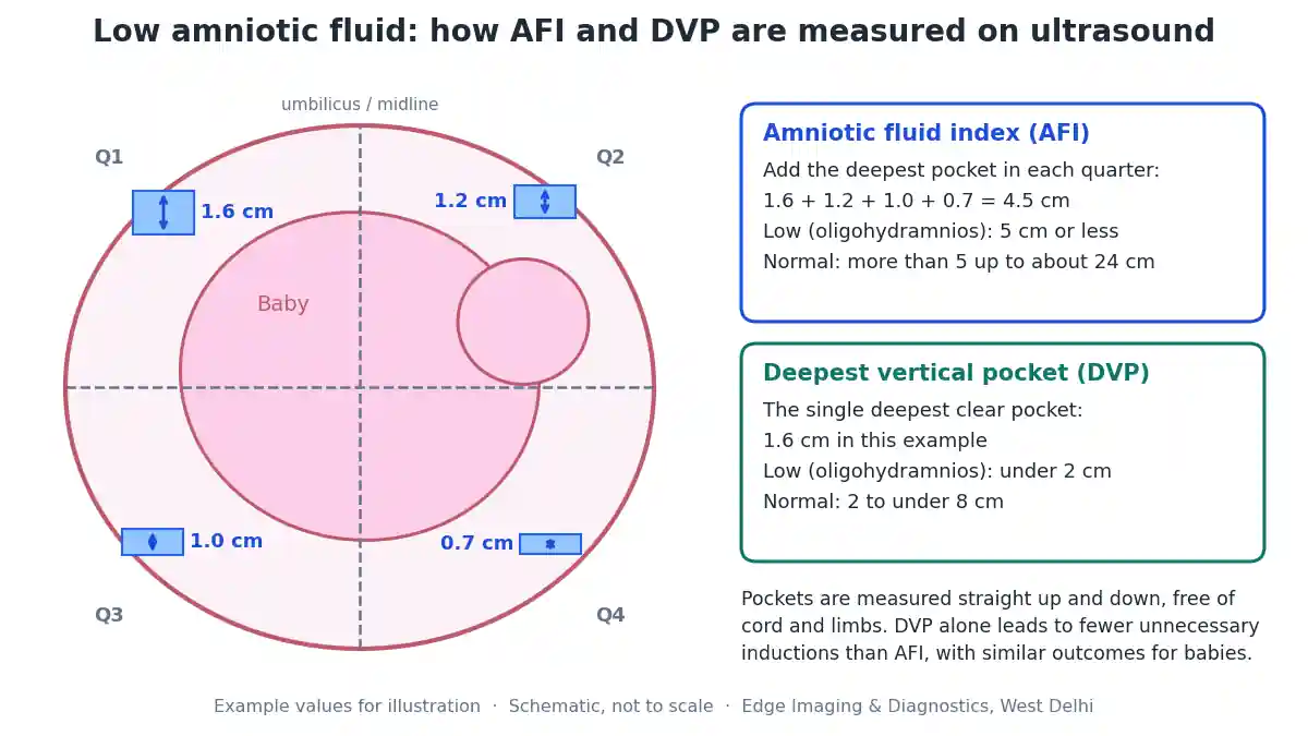
- Deepest vertical pocket (DVP), also called the single deepest pocket (SDP) or maximum vertical pocket — the depth of the single deepest clear pocket of fluid, measured straight up and down, free of cord and limbs.
- Amniotic fluid index (AFI) — the womb is divided into four quarters, the deepest pocket in each is measured, and the four numbers are added together.
| Category | Deepest vertical pocket (DVP) | Amniotic fluid index (AFI) |
|---|---|---|
| Low fluid (oligohydramnios) | Under 2 cm | 5 cm or less |
| Normal | 2 cm to under 8 cm | More than 5 cm to about 24 cm |
| Too much fluid (polyhydramnios) | 8 cm or more | About 24 to 25 cm or more |
Which measurement is better? A Cochrane review comparing the two in over three thousand women found that using the AFI led to more than twice as many diagnoses of oligohydramnios and almost twice as many inductions of labour, without any improvement in the babies’ outcomes. The reviewers concluded that the deepest vertical pocket appears to be the better choice for deciding on intervention. This is one reason why a “low AFI” on one report does not automatically mean anything needs to be done.
Borderline readings and single low measurements
Fluid measurements are not perfectly precise. The reading can change with the baby’s position, with how much pressure the probe is applied with, with whether a loop of cord was mistaken for fluid, and from one day to the next. Two operators measuring the same pregnancy on the same day can get slightly different numbers.
For this reason:
- A single borderline reading — for example an AFI of 6 or 7 cm — is usually rechecked rather than acted on.
- An AFI between about 5 and 8 cm is sometimes called “borderline”. It is not the same as oligohydramnios, and in an otherwise healthy pregnancy with normal growth it often needs only follow-up.
- What matters most is the trend over repeat scans, together with the baby’s growth, Doppler blood flow and movements.
If you have had one low reading, it is entirely reasonable to ask your doctor whether a repeat measurement, ideally using the deepest vertical pocket, is appropriate before any decision is made.
Causes of low amniotic fluid
The causes differ by the stage of pregnancy, and the cause decides the plan.
| Cause | What happens | When it is most typical |
|---|---|---|
| Rupture of the membranes | Fluid leaks out through a tear in the sac, sometimes as a slow trickle that is mistaken for urine or discharge | Any stage; early (preterm) rupture is the most important |
| Placental insufficiency | The placenta supplies less blood, so the baby diverts blood away from the kidneys and makes less urine; often linked with poor growth | Second half of pregnancy, especially with high blood pressure or pre-eclampsia |
| Post-term pregnancy | Fluid naturally falls as pregnancy goes beyond the due date | After 40 to 41 weeks |
| Kidney or urinary tract problems in the baby | Kidneys absent, small, cystic or not working, or a blockage stopping urine reaching the sac | Usually found from the second trimester |
| Certain medicines | Some painkillers (NSAIDs) and some blood-pressure tablets (ACE inhibitors and related drugs) reduce the baby’s urine | Usually second half of pregnancy |
| Chromosomal conditions | Sometimes associated with low fluid, usually with other findings | Second trimester |
| Twin complications | In identical twins sharing a placenta, one twin may have low fluid and the other too much | Second trimester |
| No cause found (idiopathic) | Fluid is low but the baby, growth and Doppler are normal | Commonest near term; outlook usually good |
Mild dehydration in the mother can also lower the reading slightly, which is one reason why drinking enough water is part of the advice given.
Symptoms: how would you know?
Most women with low amniotic fluid have no symptoms at all. It is usually discovered on a routine scan or on a scan done because the bump is measuring small.
Things that can be associated with low fluid, and that you should mention to your doctor, include:
- A gush or a steady trickle of watery fluid from the vagina, or underwear that is repeatedly damp
- A bump that seems smaller than expected for the dates
- Reduced baby movements
- Symptoms of high blood pressure or pre-eclampsia, such as headaches, swelling of the face or hands, or visual disturbance
Leaking fluid in particular should never be ignored or assumed to be urine. If you think your waters might be leaking, contact your maternity unit the same day.
Why it matters: risks at each stage of pregnancy
How serious low amniotic fluid is depends very much on when it happens and why.
| Stage | Main concerns | Typical approach |
|---|---|---|
| Second trimester (before about 24 weeks) | The baby’s lungs need fluid to grow; long-lasting severe low fluid can cause underdeveloped lungs (pulmonary hypoplasia) and stiff joints or limb position problems. A cause such as a kidney problem or early broken waters is more likely. | Detailed specialist assessment and counselling; the plan depends heavily on the cause |
| Early third trimester | Often linked with poor growth or placental problems; risk of the baby not coping well | Growth and Doppler scans, regular monitoring, steroids if early delivery is likely, carefully timed delivery |
| Near term (37 weeks onwards) | Cord compression in labour, changes in the baby’s heart rate, a higher chance of caesarean | Monitoring and, in many cases, planning delivery |
| Post-term | Fluid falls naturally; low fluid adds to the reasons to deliver | Delivery is usually advised |
In general, low fluid that is isolated — the baby is growing well, the Doppler blood flow is normal, the anatomy is normal and the waters have not broken — carries a much better outlook than low fluid that comes with poor growth, abnormal Doppler or a structural problem.
Tests after low fluid is found
Once low fluid is confirmed, the aim is to find the cause and check the baby’s wellbeing. Depending on the stage of pregnancy, your doctor may arrange:
- Checking whether the waters have broken — a speculum examination and simple bedside tests on any fluid.
- A detailed anatomy scan, with particular attention to the kidneys, bladder and urinary tract. Before about 24 weeks this is often the single most important test — see our anomaly scan (TIFFA) page.
- A growth scan with Doppler to estimate the baby’s weight and check blood flow in the umbilical cord and the baby’s brain — see our growth scan and colour Doppler guide.
- Tests of the baby’s wellbeing such as a non-stress test (heart-rate tracing) or a biophysical profile.
- The mother’s blood pressure, urine and blood tests, looking for pre-eclampsia and other conditions.
- A review of medicines taken in pregnancy.
- Genetic testing, only when other findings suggest a chromosomal or genetic condition, and only after counselling.
Occasionally, when very low fluid makes the scan too unclear to see the baby’s organs, a specialist may suggest amnioinfusion to create a fluid window for a clearer scan. In selected cases, fetal MRI can also help, because MRI pictures do not depend on fluid in the way ultrasound does.
How to increase amniotic fluid: what the evidence says
This is the question almost every family asks. Here is an honest summary of what is known.
Drinking water: modest, real, but limited
Drinking enough water is the one simple measure with research behind it. A Cochrane review of small randomised trials found that women with low fluid who drank about two litres of water had a measurable rise in their amniotic fluid index — on average about 2 cm. Intravenous fluids that are less concentrated than blood (hypotonic) also raised it, but the usual isotonic drip fluids did not.
There are important limits. The trials were small, the effect is modest and short-lived, and they did not show whether hydration improves outcomes for the baby. Drinking water will not seal broken membranes, make non-working kidneys work, or fix a struggling placenta. It is sensible advice, not a cure, and you should not drink excessive amounts. Women with heart or kidney conditions, or those told to restrict fluids, should follow their own doctor’s advice.
Rest
Lying on the left side improves blood flow to the womb, and some doctors advise extra rest. However, strict bed rest has not been shown to improve outcomes for low amniotic fluid, and it carries its own risks such as blood clots and muscle weakness. Follow your obstetrician’s individual advice.
Foods, coconut water and home remedies
Coconut water, fruit, juices and soups count towards your fluid intake, and a balanced diet is good for pregnancy. But there is no evidence that any particular food increases amniotic fluid beyond its water content. Be wary of anyone selling a product that claims otherwise.
Medicines and injections
There is currently no medicine that has been proven to safely and reliably increase amniotic fluid. Some drugs have been studied experimentally, but none is established for this purpose. Any medicine or “injection to increase fluid” offered outside a supervised specialist setting should be discussed carefully with your obstetrician before you agree to it. Conversely, if you are taking a painkiller or blood-pressure medicine that can lower fluid, your doctor may change it — never stop or change a prescribed medicine yourself.
Treating the cause
The most effective low amniotic fluid treatment is to identify and manage the underlying cause: controlling blood pressure and monitoring pre-eclampsia, managing poor growth with close surveillance, treating ruptured membranes with antibiotics and steroids where appropriate, stopping a responsible medicine, and choosing the right time for delivery.
Can a procedure or surgery increase amniotic fluid?
Many families hear that there is “a procedure” or even “surgery” to increase amniotic fluid. The honest answer has three parts.
1. Amnioinfusion can add fluid — but usually only temporarily. In amnioinfusion, warmed sterile fluid is put into the amniotic sac. Before labour this is done through a fine needle in the abdomen under ultrasound guidance (transabdominal amnioinfusion). Its main value is diagnostic: it creates a fluid window so the baby’s kidneys, bladder and other organs can be seen clearly, and it can help confirm whether the waters have broken. Because it does not fix the cause, the fluid is often lost again within hours to days. As a treatment after early broken waters, the evidence is limited and mixed. Our detailed amnioinfusion guide explains the procedure, uses and risks.
2. There is no open surgery that restores amniotic fluid. No operation on the womb permanently refills the fluid. If something is described to you as surgery to increase amniotic fluid, ask exactly what will be done, why, and what evidence supports it.
3. Specialist fetal procedures treat specific causes. In particular situations, fetal medicine specialists may treat the cause rather than the fluid — for example, placing a shunt to drain a blocked baby bladder, or laser treatment in certain identical-twin complications. Techniques to seal leaking membranes have been tried in research settings, but are not standard care. These are highly specialised decisions made in fetal medicine centres after detailed counselling.
Oligohydramnios treatment: what is done in each situation
There is no single oligohydramnios treatment. The table shows how management follows the cause.
| Situation | Usual management |
|---|---|
| Isolated low fluid, baby growing well, normal Doppler, membranes intact | Hydration advice, repeat measurement, regular monitoring; delivery planning near term |
| Low fluid with poor growth or abnormal Doppler | Close surveillance with growth, Doppler and heart-rate monitoring; steroids if early delivery is likely; delivery timed to the baby’s condition |
| Ruptured membranes before term | Hospital assessment, antibiotics, steroids where appropriate, monitoring for infection, planned timing of delivery |
| Kidney or urinary tract problem in the baby | Detailed assessment and specialist fetal medicine counselling; selected cases may be considered for a procedure |
| Medicine-related | Doctor-led review and change of medicine; fluid often recovers |
| Post-term | Delivery is usually advised |
| Very low fluid with an unclear scan | Repeat scan by an experienced operator; sometimes diagnostic amnioinfusion or fetal MRI |
Monitoring: what to expect
If you have low fluid, you can expect more frequent check-ups. Typical monitoring includes repeat fluid measurements, growth scans every two to four weeks (more often if growth is slow), Doppler studies, and heart-rate tracings or biophysical profiles once or twice a week, depending on the situation. Some women are monitored as outpatients; others, especially with broken waters or worrying findings, are admitted.
Between visits, keep track of your baby’s movements. If they become fewer or weaker than usual, contact your maternity unit the same day — do not wait for the next appointment.
Low amniotic fluid and the timing of delivery
Near the end of pregnancy, the timing of delivery is often the most important decision. For isolated, uncomplicated oligohydramnios, many experts recommend delivery at around 36 to 37 completed weeks, and if low fluid is found at 37 weeks or later, delivery is commonly advised at the time of diagnosis. The ideal timing remains debated and depends on the whole picture — the baby’s growth, Doppler results, the heart-rate tracing, whether the membranes are intact, and your own health.
Low fluid on its own does not mean you need a caesarean. Many women with low fluid have a vaginal birth with careful monitoring in labour. During labour, if the baby’s heart rate suggests the cord is being squeezed, the obstetric team has several options, which may include amnioinfusion through the cervix in the labour room. Earlier in pregnancy, when growth or Doppler is abnormal, the timing is decided case by case, balancing the risks of staying in the womb against the risks of early birth.
When the waters have broken early
When the waters break before 37 weeks and before labour, it is called preterm prelabour rupture of membranes (PPROM). It is one of the most important causes of low amniotic fluid. The main concerns are infection, early labour and, when it happens very early, the effect of prolonged low fluid on the baby’s lungs.
Management takes place in hospital and usually includes antibiotics, steroid injections to help the baby’s lungs mature when early birth is likely, close monitoring for infection, and a planned decision about when to deliver. Topping up fluid by amnioinfusion has been studied but has not been shown to clearly improve outcomes, so it is not routine.
Low fluid in twin pregnancy
In twins, fluid is measured separately in each sac, usually using the deepest vertical pocket. In identical twins who share a placenta, a striking difference — low fluid around one twin and too much around the other — can be a sign of twin-to-twin transfusion syndrome, which needs urgent specialist assessment. This is one of the reasons identical twin pregnancies are scanned more frequently.
The outlook for the baby
The outlook varies widely, and it is driven by the cause rather than the fluid number.
- Isolated low fluid near term, with normal growth and Doppler, generally has a good outcome with monitoring and well-timed delivery.
- Low fluid with poor growth reflects the placental problem; the outlook depends on how early it occurs and how well the baby is monitored and delivered.
- Severe low fluid from early in the second trimester, especially from very early broken waters or serious kidney problems, carries a more guarded outlook because of the effect on lung development. Specialist counselling is essential in this situation.
Every pregnancy is different. Your obstetrician and fetal medicine specialist can give you an outlook that reflects your own scans and circumstances.
Myths and facts
| Myth | Fact |
|---|---|
| A single low AFI means the baby is in danger | One reading can be misleading; the trend, the cause and the baby’s growth and Doppler matter far more |
| Coconut water or special foods increase fluid | Only their water content helps; no food has a special effect |
| An injection can fix low fluid | No medicine is proven to safely and reliably raise amniotic fluid |
| Surgery can refill the fluid permanently | There is no such surgery; amnioinfusion adds fluid temporarily, mainly for diagnosis |
| Low fluid always means a caesarean | Many women deliver vaginally with careful monitoring |
| Drinking huge amounts of water is better | Adequate hydration helps modestly; excessive intake is not recommended |
When to seek help urgently
Contact your maternity unit or go to hospital promptly if you have:
- A gush or steady leak of watery fluid from the vagina
- Reduced, weaker or absent baby movements
- Vaginal bleeding
- Fever, shivering or offensive vaginal discharge
- Regular tightenings or contractions before 37 weeks
- Severe headache, visual disturbance, or sudden swelling of the face or hands
- Severe or persistent abdominal pain
Questions to ask your doctor
- Was my fluid measured by AFI or deepest vertical pocket, and what were the numbers?
- Should the measurement be repeated before we decide anything?
- What do you think is the cause in my case?
- Is my baby growing normally, and is the Doppler normal?
- Have my waters broken?
- Are any of my medicines a possible cause?
- How often will I be monitored, and where?
- Would a detailed anomaly scan, amnioinfusion or fetal MRI add anything in my case?
- When do you think delivery should be planned, and why?
Prenatal scans and the law
Prenatal ultrasound and diagnostic procedures in India are governed by the Pre-Conception and Pre-Natal Diagnostic Techniques (PCPNDT) Act. Scans are performed to assess the baby’s health and development at a registered centre. Determining or disclosing the sex of a fetus is a criminal offence. We do not determine it and do not disclose it under any circumstances.
Low amniotic fluid scans in Delhi
Edge Imaging & Diagnostics is a NABH-accredited diagnostic group in West Delhi, registered under the PCPNDT Act. For women with low amniotic fluid we offer the scans that matter — amniotic fluid assessment (AFI and deepest vertical pocket), growth scans with colour Doppler, detailed anomaly scans (TIFFA), fetal echocardiography and fetal MRI — and, on an obstetrician’s referral, ultrasound-guided procedures including transabdominal amnioinfusion and amniocentesis.
Reports are prepared by our consultant radiologists and sent to your obstetrician, who plans your care. If a repeat measurement or a detailed scan is the right next step rather than a procedure, we will tell you that plainly.
Send a photograph of your latest scan report or your doctor’s prescription on WhatsApp and we will tell you which scan is being asked for, the preparation, and today’s actual pricing.
💬 Send your report on WhatsApp for guidance and today’s pricing | Call 9555999960
Related reading: Amnioinfusion · Too much fluid: amnioreduction for polyhydramnios · Growth scan and Doppler · Anomaly scan (TIFFA) · Fetal MRI · Pregnancy scan schedule
References
- Oligohydramnios — StatPearls, NCBI Bookshelf (US National Library of Medicine).
- Oligohydramnios — Merck Manual, Professional Edition.
- Maternal hydration for increasing amniotic fluid volume in oligohydramnios and normal amniotic fluid volume — Cochrane Database of Systematic Reviews.
- Amniotic fluid index compared with single deepest vertical pocket measurement in predicting an adverse pregnancy outcome — Cochrane.
- Government of NCT of Delhi: PCPNDT — Delhi.
Frequently asked questions
What is low amniotic fluid?
Low amniotic fluid, or oligohydramnios, means there is less fluid around the baby than expected for the stage of pregnancy. It is usually diagnosed on ultrasound when the deepest vertical pocket is under 2 cm or the amniotic fluid index is 5 cm or less.
What is a normal AFI in pregnancy?
An amniotic fluid index of more than 5 cm up to about 24 cm is generally considered normal. A deepest vertical pocket of 2 cm to under 8 cm is normal. Values change with the stage of pregnancy, so your report’s reference range and your doctor’s interpretation matter.
What causes low amniotic fluid?
Common causes include leaking or ruptured membranes, a placenta that is not supplying the baby well (often with poor growth), pregnancy beyond the due date, kidney or urinary tract problems in the baby, and certain medicines such as NSAID painkillers and ACE-inhibitor blood-pressure tablets. Sometimes no cause is found.
Is low amniotic fluid dangerous for the baby?
It depends on the cause and the stage of pregnancy. Isolated low fluid near term with normal growth usually has a good outcome with monitoring and well-timed delivery. Severe low fluid early in the second trimester, or low fluid with poor growth, is more serious and needs specialist care.
How can I increase amniotic fluid naturally?
Drinking enough water is the one simple measure with evidence: in small trials, drinking about two litres raised the amniotic fluid index by around 2 cm on average. It does not treat underlying causes, and no food or supplement has a special effect beyond its water content.
Is there an injection or medicine to increase amniotic fluid?
No medicine has been proven to safely and reliably increase amniotic fluid. Some drugs have been studied experimentally, but none is established. Discuss any medicine or injection offered for low fluid with your obstetrician before agreeing to it.
Is there a procedure or surgery to increase amniotic fluid?
There is no surgery that permanently restores amniotic fluid. Amnioinfusion can add warmed sterile fluid to the sac through a fine needle under ultrasound, mainly to get a clear scan and find the cause, but the fluid is often lost again because the cause is not fixed.
Does drinking coconut water increase amniotic fluid?
Coconut water counts towards your daily fluid intake, which can help modestly, but there is no evidence that it has any special effect on amniotic fluid compared with plain water.
Can low amniotic fluid become normal again?
Yes, sometimes. Fluid can improve when a cause such as dehydration or a medicine is corrected, and a borderline reading may be normal on a repeat scan. When the cause is ruptured membranes, placental insufficiency or a kidney problem, the fluid usually stays low.
Does low amniotic fluid mean I need a caesarean?
Not necessarily. Many women with low fluid have a vaginal birth with careful monitoring in labour. A caesarean is advised when there are other reasons, such as concerns about the baby’s heart rate, growth or position.
When is delivery advised for low amniotic fluid?
For isolated, uncomplicated low fluid, many experts recommend delivery at around 36 to 37 completed weeks, and if it is found at 37 weeks or later, delivery is commonly advised at diagnosis. With poor growth or abnormal Doppler, timing is decided case by case.
Which is better, AFI or deepest vertical pocket?
A Cochrane review found that using the AFI diagnosed low fluid more often and led to more inductions of labour without improving babies’ outcomes, so the deepest vertical pocket appears to be the better measurement for deciding on intervention.
Can low amniotic fluid affect the baby’s lungs?
Yes, when severe low fluid lasts for a long time in the second trimester, roughly before 24 weeks, it can prevent the lungs from growing properly. Low fluid later in pregnancy does not have this effect on lung growth.
What symptoms should I watch for with low amniotic fluid?
Seek prompt assessment for leaking fluid from the vagina, reduced baby movements, bleeding, fever, contractions before 37 weeks, severe headache or visual disturbance, or severe abdominal pain.
Which scans are done for low amniotic fluid?
Typically repeat fluid measurement, a growth scan with colour Doppler, and a detailed anomaly scan focusing on the kidneys and bladder. Heart-rate monitoring or a biophysical profile checks the baby’s wellbeing. Fetal MRI or diagnostic amnioinfusion is occasionally used when the ultrasound view is poor.
Does bed rest help low amniotic fluid?
Resting on the left side can improve blood flow to the womb, but strict bed rest has not been shown to improve outcomes and carries risks such as blood clots. Follow your obstetrician’s individual advice.
Where can I get a low amniotic fluid scan in Delhi?
Edge Imaging & Diagnostics in West Delhi offers amniotic fluid assessment, growth scans with colour Doppler, anomaly scans and fetal MRI, with reports sent to your obstetrician. Send your report or prescription on WhatsApp to 9555999960 for guidance and today’s pricing.
The bottom line
Low amniotic fluid is a scan finding, not a diagnosis in itself. What matters is why it is low, how far along you are, and whether the baby is growing and coping well. One borderline reading often needs only a repeat measurement.
Drinking enough water helps modestly. No medicine or surgery permanently restores fluid; amnioinfusion adds fluid temporarily, mainly to get a clear scan. Careful monitoring and well-timed delivery remain the most effective tools.
Written by the clinical content team at Edge Imaging & Diagnostics, West Delhi — a NABH-accredited diagnostic centre registered under the PCPNDT Act. Medically reviewed by Dr. Nishant Thapar, Consultant Radiologist. Last reviewed: October 2026.
Medical disclaimer: this article is general information about low amniotic fluid. It is not medical advice, a diagnosis, or guidance on any decision about a pregnancy.
Measurement cut-offs, effect sizes and delivery timings quoted here are approximate and vary between guidelines, centres and individual pregnancies. Your own obstetrician should give you advice specific to your situation.
Do not take any medicine or supplement for low amniotic fluid, and do not stop a prescribed medicine, without your doctor’s advice. Leaking fluid, reduced baby movements, bleeding, fever or pain need prompt in-person assessment.
Prenatal ultrasound in India is governed by the PCPNDT Act. Determining or disclosing the sex of a fetus is a criminal offence, and this centre does not do so under any circumstances.