
 PV
 TG
 MN
 EM
 EI
 ND

 CT Scan
 PET CT
 Digital X-ray
 Ultrasound
 Amniocentesis

 Fetal Reduction
 Thermalytix Breast Screening
 Cardiology
 Lab Tests
 GAMCA Pre-Medical

Amnioreduction is a procedure that removes excess amniotic fluid from around the baby, through a fine needle passed into the womb under continuous ultrasound guidance. It is used when there is far too much fluid — a condition called polyhydramnios — and that fluid is causing the mother significant breathlessness or pain, or raising the risk of early labour.
If you have been told your scan shows “excess fluid” or “polyhydramnios”, the first thing to know is that most cases are mild, need no procedure at all, and are simply watched with repeat scans. Amnioreduction is for the smaller group in whom the fluid is severe and genuinely troublesome. This guide explains how amniotic fluid is measured, what causes too much of it, when amnioreduction is the right step, exactly how it is done, the honest risks, and why the fluid often comes back.
It is general information, not a decision. That belongs to you and your obstetrician.
Amnioreduction at a glance
- What it is: drainage of excess amniotic fluid through a fine needle, under ultrasound guidance
- Why: severe polyhydramnios causing maternal breathlessness, pain or tightening, or raising the risk of preterm labour; also some twin complications
- When: usually in the second half of pregnancy, when symptoms or risk justify it
- How long: roughly half an hour to an hour, because fluid is removed slowly
- Anaesthesia: local anaesthetic in the skin; no general anaesthesia
- Main limitation: it treats the excess fluid, not its cause — the fluid often re-accumulates and the procedure may need repeating
- Main risks: leaking of fluid or rupture of membranes, preterm labour, infection and, rarely, placental abruption
What this guide covers
- What polyhydramnios (too much amniotic fluid) means
- How amniotic fluid is measured: AFI and DVP
- What causes excess amniotic fluid
- Symptoms of polyhydramnios
- Why too much fluid matters
- Finding the cause comes first
- When amnioreduction is needed — and when it is not
- Amnioreduction in twin pregnancy and TTTS
- Other options your doctor may discuss
- Before the procedure
- How amnioreduction is done, step by step
- Does amnioreduction hurt?
- Amnioreduction risks: the honest picture
- After the procedure
- Why the fluid often comes back
- Polyhydramnios and delivery
- Polyhydramnios treatment options compared
- Amnioreduction vs amniocentesis
- What to expect on the day
- Amnioreduction in Delhi: where it is done
- References
- Frequently asked questions
What polyhydramnios (too much amniotic fluid) means
The baby floats in amniotic fluid throughout pregnancy. That fluid cushions the baby, lets it move and stretch its limbs, helps the lungs develop, and keeps the umbilical cord from being squeezed. Its volume is kept in balance by two processes: the baby passes urine into it, and the baby swallows it.
Polyhydramnios means there is more fluid than the normal range for that stage of pregnancy. It happens in roughly one to two pregnancies in every hundred. When the balance tips towards too much fluid, it is usually because the baby is producing more urine than usual, or is swallowing less than usual, or because of a maternal factor such as diabetes. In a large proportion of mild cases, no cause is found at all.
The opposite problem — too little fluid — is called oligohydramnios and is managed quite differently; one of the procedures used there is explained in our guide to amnioinfusion.
How amniotic fluid is measured: AFI and DVP
Amniotic fluid cannot be measured directly by volume on a scan, so two standard ultrasound measurements are used instead. Your report will state which one was used.
- Deepest vertical pocket (DVP), also called the single deepest pocket (SDP) — the depth of the single deepest clear pocket of fluid, measured straight up and down, in centimetres.
- Amniotic fluid index (AFI) — the womb is divided into four quarters, the deepest pocket in each is measured, and the four numbers are added together, in centimetres.
Commonly used ranges are shown below. Exact cut-offs differ slightly between guidelines, so always read your own report’s reference range.
| Category | Deepest vertical pocket (DVP) | Amniotic fluid index (AFI) |
|---|---|---|
| Low fluid (oligohydramnios) | Under 2 cm | Under 5 cm |
| Normal | 2 to 7.9 cm | 5 to 24.9 cm |
| Mild polyhydramnios | 8 to 11.9 cm | 25 to 29.9 cm |
| Moderate polyhydramnios | 12 to 15.9 cm | 30 to 34.9 cm |
| Severe polyhydramnios | 16 cm or more | 35 cm or more |
Two practical points. First, a single borderline measurement is not a diagnosis to panic about — fluid measurements vary with the baby’s position and between scans, and a repeat measurement is often the sensible next step. Second, the severity category matters a great deal: mild polyhydramnios is common and usually harmless, while severe polyhydramnios is more likely to have a cause and to cause problems.
What causes excess amniotic fluid
- No identifiable cause (idiopathic) — the single commonest situation, especially in mild cases. The baby is otherwise well, and the outlook is generally good.
- Diabetes in the mother — pre-existing or gestational. Higher blood sugar in the baby increases urine production. This is one of the most important treatable causes, and good sugar control often reduces the fluid.
- Problems with the baby’s swallowing or gut — for example a blockage of the food pipe or the upper intestine, which stops swallowed fluid moving on. These are usually looked for carefully on a detailed anomaly scan.
- Conditions affecting the baby’s muscles or nervous system that reduce swallowing.
- Twin-to-twin transfusion syndrome in identical twins who share one placenta, where one twin has far too much fluid and the other far too little.
- Fetal anemia or heart failure, sometimes with fluid collecting in the baby’s body (hydrops).
- Infections passed from mother to baby.
- Chromosomal and genetic conditions, usually alongside other findings on the scan.
- Rare kidney conditions in the baby that cause very high urine output.
The more severe the polyhydramnios, and the earlier in pregnancy it appears, the more likely it is that a specific cause will be found.
Symptoms of polyhydramnios
Mild polyhydramnios often causes no symptoms and is found only on a routine scan. As the fluid increases, the womb becomes larger and tighter than expected, and women may notice:
- A bump that is much larger than expected for the dates, or that has grown quickly
- Breathlessness, especially lying flat, because the enlarged womb pushes up on the diaphragm
- A tight, stretched, uncomfortable abdomen, sometimes painful
- Indigestion, heartburn or reduced appetite from pressure on the stomach
- Swelling of the legs and passing urine less often than usual
- Tightenings or contractions before term
- Difficulty feeling the baby’s movements, because the baby has so much room
Seek prompt assessment if you have regular contractions before 37 weeks, a gush or steady leak of fluid, bleeding, severe abdominal pain, or breathlessness at rest. Sudden severe breathlessness or chest pain is an emergency.
Why too much fluid matters
Excess fluid stretches the womb, and an over-stretched womb is more likely to contract early. Polyhydramnios is associated with a higher chance of:
- Preterm labour and early rupture of the membranes
- Malpresentation — the baby lying sideways or breech, because there is so much room to turn
- Cord prolapse when the waters break, because a rush of fluid can carry the cord ahead of the baby
- Placental abruption if a large volume of fluid is lost suddenly
- Heavy bleeding after delivery, because an over-stretched womb contracts less efficiently
These risks are much higher with severe polyhydramnios than with mild. They explain both why it is monitored and why, in selected cases, the fluid is reduced.
Finding the cause comes first
Before anyone considers draining fluid, the more important task is understanding why it is there. A typical assessment includes:
- A detailed anomaly scan of the baby, looking particularly at the stomach, gut, chest, brain, limbs and heart. See our anomaly scan (TIFFA) guide.
- Fetal echocardiography where the heart needs a closer look — see fetal echocardiography in Delhi.
- Doppler studies, including the middle cerebral artery Doppler if fetal anemia is a possibility — see our growth scan and Doppler guide and our guide to cordocentesis for suspected fetal anemia.
- A test for diabetes, usually a glucose tolerance test, arranged by your doctor.
- Blood tests for infection, where appropriate.
- Genetic testing when the scan shows additional findings. This can be done by amniocentesis — and conveniently, if an amnioreduction is being done anyway, a sample of the removed fluid can be sent for testing at the same time.
When amnioreduction is needed — and when it is not
Amnioreduction is not needed for most polyhydramnios. Mild and moderate cases without troublesome symptoms are usually managed by treating any cause (such as tightening diabetes control) and monitoring with repeat scans.
It is generally considered when:
- Polyhydramnios is severe, and
- it is causing significant maternal symptoms — breathlessness, pain, inability to lie down or eat — or
- it is causing preterm contractions or a short cervix, where reducing the stretch may help prolong the pregnancy, or
- it is part of a twin complication such as twin-to-twin transfusion syndrome, in specific circumstances.
Your specialist weighs the benefit — relief of symptoms and possibly a longer pregnancy — against the small risks of the procedure itself. Where the fluid is mild and you are comfortable, the balance almost always favours simply watching.
Amnioreduction in twin pregnancy and TTTS
Identical twins who share one placenta (monochorionic twins) share blood vessels across that placenta. If blood flows unevenly between them, twin-to-twin transfusion syndrome (TTTS) can develop: the receiving twin produces large amounts of urine and is surrounded by excess fluid, while the donor twin has very little.
For the more advanced stages of TTTS in mid-pregnancy, the treatment of choice in specialist centres is generally fetoscopic laser treatment, which seals the connecting vessels and treats the cause. Amnioreduction, which only removes the excess fluid from the recipient’s sac, may still be used in particular situations — for example later in pregnancy, at an early stage, or where laser treatment is not possible or appropriate. If you have monochorionic twins, regular scans from early in pregnancy are what catch TTTS in time, and your fetal medicine specialist will explain which treatment fits your situation.
Other options your doctor may discuss
- Treating the cause — above all, tight blood sugar control in diabetes, which can reduce fluid by itself.
- Watchful monitoring with repeat scans, for mild and moderate cases.
- Medication — in some situations, and only before a certain stage of pregnancy, specialists may consider a medicine that reduces the baby’s urine output. It carries specific risks for the baby’s heart circulation and must only be used under close specialist supervision. It is never something to take on your own.
- Planning delivery at an appropriate time and place, which for many women is the most important part of management.
Before the procedure
- A counselling conversation covering why it is recommended, what it can and cannot achieve, the risks, the likelihood of needing it again, and the alternatives.
- Written consent. If any fluid is to be sent for genetic testing, the documentation required under the PCPNDT Act is also completed.
- Your blood group. Rh-negative mothers who are not already sensitised are usually given anti-D afterwards.
- Your medicines, especially blood thinners. Tell the team about everything; do not stop anything on your own.
- Previous reports — earlier fluid measurements, anomaly scan, Doppler and any sugar test results.
- Someone to accompany you and take you home.
How amnioreduction is done, step by step
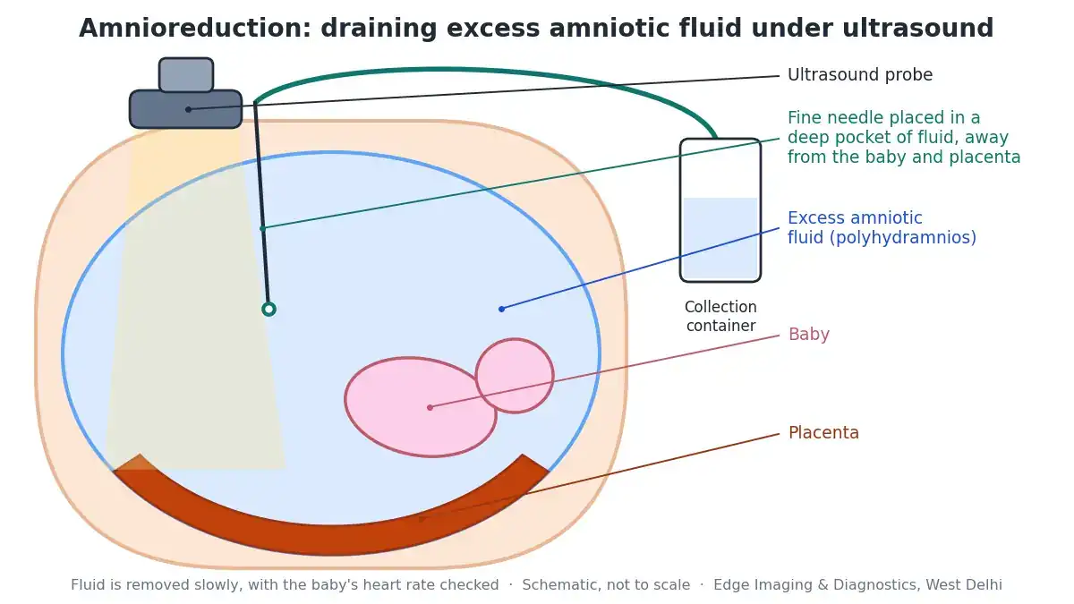
- Scan first. An ultrasound confirms the baby’s position and heartbeat, the placenta, the cervix if relevant, and the deepest pocket of fluid well away from the baby and the placenta.
- Cleaning and numbing. The abdomen is cleaned with antiseptic, covered with sterile drapes, and the skin is numbed with local anaesthetic.
- Needle placement. A fine needle is guided into the chosen pocket while the specialist watches on the screen.
- Slow drainage. The fluid is drained through tubing into a collection container, often with gentle suction. It is removed slowly — usually over roughly half an hour to an hour — because a rapid drop in pressure inside the womb increases the risk of complications. Commonly a litre or more is removed, sometimes considerably more, depending on how much is present.
- Monitoring throughout. The baby’s heart rate and the needle position are checked repeatedly. The drainage is stopped when the fluid measurement approaches the upper part of the normal range, or earlier if needed.
- Samples if needed. A small amount of the fluid can be sent for genetic or infection testing if that has been requested.
- Finish. The needle is removed, and the baby’s heart rate and the placenta are checked again.
Does amnioreduction hurt?
Most women feel a sharp sting as the local anaesthetic goes in, then pressure, and sometimes cramping during the drainage. Because the procedure takes longer than an amniocentesis, lying still is the main discomfort. Many women notice immediate relief of breathlessness and tightness as the womb softens, which is often the first sign that it has worked.
Amnioreduction risks: the honest picture
Amnioreduction is generally considered a low-risk procedure in experienced hands, but it is not risk-free, and the risks need to be weighed against the risks of severe polyhydramnios itself. Published series report procedure-related complications in a small percentage of procedures. They include:
- Leaking of fluid or rupture of the membranes
- Preterm labour — although in some women reducing the fluid actually helps prolong the pregnancy
- Placental abruption — separation of the placenta, which is the main reason fluid is drained slowly; uncommon, but serious
- Infection inside the womb, which is rare with sterile technique
- A temporary change in the baby’s heart rate during the procedure
- Rh sensitisation in Rh-negative mothers, prevented by anti-D
The risk is higher with repeated procedures and in complex twin pregnancies. Ask your specialist for their own experience and complication figures — it is a reasonable question, and a good specialist answers it readily.
After the procedure
- You will be monitored for a while afterwards, and the baby’s heart rate is checked before you leave. In some situations, particularly with contractions or later in pregnancy, a period of observation in hospital is advised.
- Rest for the remainder of the day and avoid heavy lifting or strenuous activity for a day or two, or as advised.
- Mild cramping and a little leakage at the needle site can occur.
Contact your doctor or go to hospital promptly for regular contractions, a gush or steady leak of fluid, bleeding, fever or chills, severe abdominal pain, or reduced baby movements.
Why the fluid often comes back
This is the part most families are not told in advance. Amnioreduction removes the excess fluid; it does not usually remove the reason the fluid built up. If the baby continues to produce more fluid than it swallows, the fluid gradually re-accumulates — sometimes over days, sometimes over weeks.
That means some women need more than one amnioreduction during a pregnancy. The interval is guided by repeat scans and, above all, by symptoms. Where the cause can be treated — diabetes being the clearest example — the fluid may settle without further procedures.
Polyhydramnios and delivery
- Plan to deliver in a hospital with full obstetric and newborn facilities, particularly if a cause has been found in the baby.
- When your waters break, go to hospital straight away. With a large volume of fluid, there is a higher chance of the cord slipping down ahead of the baby (cord prolapse), which is an emergency.
- The baby’s position is checked near term, because babies in a lot of fluid are more likely to lie sideways or breech.
- The team will be prepared for heavier bleeding after birth, which is more common when the womb has been over-stretched.
- After birth, the newborn team may examine the baby specifically for the cause suspected in pregnancy — for example by checking that a feeding tube passes into the stomach.
With mild, unexplained polyhydramnios, most babies are born healthy and need nothing more than a routine newborn check.
Polyhydramnios treatment options compared
Polyhydramnios treatment is a ladder rather than a single step. Most women only ever need the first two rungs. The table below sets the options side by side.
| Option | When it is used | What it does | Limits |
|---|---|---|---|
| Find and treat the cause | Every case | Detailed scans, a diabetes test and other tests identify why the fluid is high; treating diabetes can reduce fluid by itself | In many mild cases no cause is found |
| Monitoring with repeat scans | Mild and moderate polyhydramnios without troublesome symptoms | Tracks fluid, growth and the baby’s wellbeing at intervals | Does not reduce the fluid; relies on symptoms being tolerable |
| Medication under specialist care | Selected cases, only before a certain stage of pregnancy | Reduces the baby’s urine output | Specific risks to the baby’s heart circulation; needs close monitoring |
| Amnioreduction | Severe, symptomatic polyhydramnios or preterm risk; some twin complications | Drains excess fluid; often gives immediate relief | Fluid may return; small procedure risks |
| Fetoscopic laser | Advanced twin-to-twin transfusion syndrome in mid-pregnancy, at specialist centres | Seals the shared vessels, treating the cause in TTTS | Only for TTTS; specialist availability |
| Delivery planning | Every case | Right hospital, right timing, readiness for cord prolapse and bleeding | — |
Amnioreduction vs amniocentesis: what is the difference?
The two are often confused because they look almost identical from the outside: both use a fine needle passed into the womb under ultrasound guidance. The difference is in the purpose and the volume.
- Amniocentesis is diagnostic. A small sample — typically around 15 to 20 millilitres — is taken to test the baby’s chromosomes, genes or for infection. It takes a minute or two.
- Amnioreduction is therapeutic. A large volume — often a litre or more — is removed to relieve the effects of excess fluid. Because the fluid must be drained slowly, it takes much longer, usually half an hour to an hour.
- They can be combined. If testing is needed and an amnioreduction is being done anyway, a sample of the drained fluid can be sent to the laboratory, so a separate amniocentesis is not required.
For the diagnostic test itself, see our amniocentesis guide.
What to expect on the day of the amnioreduction procedure
- Arrival and review. Your referral, previous scans and blood group are checked, and you have a chance to ask questions before anything begins.
- Consent. The reasons, risks, the chance of needing a repeat, and the alternatives are explained, and consent forms are signed.
- Baseline scan. The fluid is measured, the baby’s heartbeat and position are checked, and the safest pocket is chosen.
- The amnioreduction procedure itself. Usually around half an hour to an hour, with regular checks of the baby’s heart rate throughout.
- Post-procedure scan. The fluid is re-measured and the baby and placenta are checked again.
- Observation. You rest for a period before going home, or are observed in hospital if your obstetrician has advised it, for example if you were having contractions.
- Follow-up plan. You leave knowing when your next scan is due and which symptoms should bring you back sooner.
Allow a good part of the day, and do not plan to drive yourself home.
Prenatal procedures and the law
Prenatal diagnostic procedures in India are governed by the Pre-Conception and Pre-Natal Diagnostic Techniques (PCPNDT) Act. Any testing of amniotic fluid is done only to detect genetic, chromosomal, metabolic or structural conditions, at a registered centre. Determining or disclosing the sex of a fetus is a criminal offence. We do not determine it and do not disclose it under any circumstances.
Amnioreduction in Delhi: where it is done
Edge Imaging & Diagnostics is a NABH-accredited diagnostic group in West Delhi, registered under the PCPNDT Act, offering ultrasound-guided amnioreduction in Delhi together with the scans that matter in polyhydramnios — amniotic fluid assessment, anomaly scans, fetal Doppler and fetal echocardiography — and related procedures such as amniocentesis. Procedures are performed by our consultant radiologists on an obstetrician’s referral.
Before anything is booked, we review your referral and your recent scans. If the fluid is mild and a repeat scan is the right next step rather than a procedure, we will tell you that plainly.
Send a photograph of your obstetrician’s referral and your latest scan report on WhatsApp and we will explain what is being asked for, the preparation, and today’s actual pricing.
💬 Send your referral on WhatsApp for guidance and today’s pricing | Call 9555999960
Related reading: Amniocentesis in Delhi · Cordocentesis (fetal blood sampling) · Anomaly scan (TIFFA) · Growth scan and Doppler · Fetal echocardiography · Pregnancy scan schedule
References
- Polyhydramnios — StatPearls, NCBI Bookshelf (US National Library of Medicine).
- Polyhydramnios: Causes, Diagnosis and Therapy — PubMed Central.
- Amnioreduction for Polyhydramnios in a Consecutive Series at a Single Center: Indications, Risks and Perinatal Outcomes — Children, 2024.
- Too Much of a Good Thing: Updated Current Management and Perinatal Outcomes of Polyhydramnios — PubMed.
- Government of NCT of Delhi: PCPNDT — Delhi.
Frequently asked questions
What is amnioreduction?
Amnioreduction is a procedure that removes excess amniotic fluid from around the baby through a fine needle passed into the womb under continuous ultrasound guidance. It is used for severe polyhydramnios that is causing significant symptoms or raising the risk of preterm labour, and in some twin complications.
What is polyhydramnios?
Polyhydramnios means there is more amniotic fluid than normal for the stage of pregnancy. It is usually defined as a deepest vertical pocket of 8 cm or more, or an amniotic fluid index of about 24 to 25 cm or more. It occurs in roughly one to two pregnancies in a hundred, and most cases are mild.
Is polyhydramnios dangerous?
Mild polyhydramnios with no identifiable cause usually has a good outcome. Severe polyhydramnios is more likely to have an underlying cause and is associated with preterm labour, malpresentation, cord prolapse when the waters break, placental abruption and heavier bleeding after birth, which is why it is monitored closely.
Does every case of excess amniotic fluid need amnioreduction?
No. Most polyhydramnios, especially mild and moderate, is managed by treating any cause such as diabetes and by monitoring with repeat scans. Amnioreduction is generally reserved for severe polyhydramnios causing significant breathlessness, pain or preterm contractions, or for specific twin complications.
How much fluid is removed during amnioreduction?
It varies with how much is present. Commonly a litre or more is removed, sometimes considerably more, drained slowly over roughly half an hour to an hour. Drainage usually stops when the fluid measurement approaches the upper part of the normal range.
Is amnioreduction painful?
Most women feel a sharp sting with the local anaesthetic, then pressure and sometimes cramping during drainage. General anaesthesia is not used. Many women notice immediate relief of breathlessness and abdominal tightness as the fluid is removed.
What are the risks of amnioreduction?
Published series report complications in a small percentage of procedures, including leaking of fluid or rupture of the membranes, preterm labour, placental abruption, infection, and temporary changes in the baby’s heart rate. Fluid is drained slowly specifically to reduce the risk of abruption. Risks are higher with repeated procedures and in complex twin pregnancies.
Will the fluid come back after amnioreduction?
Often, yes. Amnioreduction removes the excess fluid but usually not its cause, so fluid can re-accumulate over days or weeks and the procedure may need repeating. Where a cause such as diabetes can be treated, the fluid may settle without further procedures.
What causes too much amniotic fluid?
Often no cause is found, particularly in mild cases. Known causes include maternal diabetes, conditions that reduce the baby’s swallowing or block its gut, twin-to-twin transfusion syndrome, fetal anemia, infections, chromosomal conditions and rare fetal kidney conditions.
How is amniotic fluid measured on ultrasound?
Either as the deepest vertical pocket (DVP), the depth of the single deepest pocket of fluid, or as the amniotic fluid index (AFI), the sum of the deepest pockets in the four quarters of the womb. Your report states which method was used and the reference range applied.
Can diabetes cause excess amniotic fluid?
Yes. High blood sugar in the mother leads to higher sugar levels in the baby, which increases the baby’s urine output. Diabetes is one of the most important treatable causes of polyhydramnios, and improving sugar control can reduce the fluid.
What is the role of amnioreduction in twin-to-twin transfusion syndrome?
In advanced mid-pregnancy TTTS, fetoscopic laser treatment that seals the connecting vessels is generally the treatment of choice in specialist centres because it treats the cause. Amnioreduction, which only removes the recipient twin’s excess fluid, may be used in particular situations such as later in pregnancy or early-stage disease. Your fetal medicine specialist will advise.
Can the removed fluid be tested?
Yes. If genetic or infection testing is needed, a sample of the drained fluid can be sent to the laboratory at the same time, avoiding a separate amniocentesis. This is done within the PCPNDT Act and never to determine the sex of the baby.
What should I do if my waters break with polyhydramnios?
Go to hospital straight away. With a large volume of fluid, there is a higher chance of the umbilical cord slipping down ahead of the baby, which is an emergency. Plan to deliver in a hospital with full obstetric and newborn facilities.
What symptoms should I watch for with excess fluid?
Seek prompt assessment for regular contractions before 37 weeks, a gush or steady leak of fluid, bleeding, severe abdominal pain, fever, reduced baby movements, or breathlessness at rest. Sudden severe breathlessness or chest pain is an emergency.
Is amnioreduction available in Delhi?
Yes. Amnioreduction in Delhi is performed at fetal medicine and imaging centres on an obstetrician’s referral. Edge Imaging & Diagnostics in West Delhi performs ultrasound-guided amnioreduction along with anomaly scans, fetal Doppler and fetal echocardiography. Send your referral on WhatsApp to 9555999960.
Do I need a referral for amnioreduction?
Yes. It is performed on an obstetrician’s or fetal medicine specialist’s referral after the cause of the excess fluid has been assessed, and with consent covering the risks, the likelihood of repeat procedures and the alternatives.
The bottom line
Too much amniotic fluid is usually mild, often unexplained, and usually just watched. When it is severe — making you breathless, painful or contracting early — amnioreduction drains the excess slowly through a fine needle under ultrasound, and many women feel relief straight away.
It treats the fluid rather than its cause, so the fluid can come back and the procedure may need repeating. Finding and treating the cause, and planning a safe delivery, matter just as much as the procedure itself.
Written by the clinical content team at Edge Imaging & Diagnostics, West Delhi — a NABH-accredited diagnostic centre registered under the PCPNDT Act. Medically reviewed by Dr. Nishant Thapar, Consultant Radiologist. Last reviewed: October 2026.
Medical disclaimer: this article is general information about polyhydramnios and amnioreduction. It is not medical advice, a diagnosis, or guidance on any decision about a pregnancy.
Fluid measurement cut-offs, volumes and risk figures quoted here are approximate and vary between guidelines, centres and individual pregnancies. Your own obstetrician and the specialist performing the procedure should give you figures specific to your situation.
Do not take any medicine to reduce amniotic fluid except under specialist supervision. Contractions, leaking fluid, bleeding, fever, severe pain, reduced fetal movements or breathlessness at rest need prompt in-person assessment.
Prenatal diagnostic testing in India is governed by the PCPNDT Act. Determining or disclosing the sex of a fetus is a criminal offence, and this centre does not do so under any circumstances.