
 PV
 TG
 MN
 EM
 EI
 ND
 NM
 SR

 CT Scan
 PET CT
 Digital X-ray
 Ultrasound
 Amniocentesis

 Fetal Reduction
 Thermalytix Breast Screening
 Cardiology
 Lab Tests
 GAMCA Pre-Medical

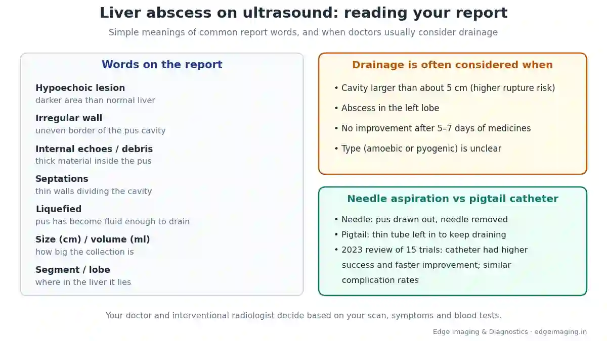
A liver abscess is a collection of pus inside the liver. On ultrasound it usually appears as a darker (hypoechoic) area with an irregular wall, sometimes with debris or septations inside. The report’s size, liquefaction and location help your doctor decide between medicines alone, needle aspiration or a pigtail catheter drain.
You have just collected an ultrasound report. Somewhere in the middle are words like “hypoechoic lesion in segment VII, 6.2 × 5.8 cm, approximately 110 ml, with internal echoes — likely liver abscess, partially liquefied.” Your doctor has mentioned drainage. Your family is worried. And the report reads like a foreign language.
This guide translates that report into simple words. It explains what each common term means, what the size and volume tell your doctor, when drainage is usually considered, and the difference between needle aspiration and a pigtail catheter. It is written for patients and families — not for doctors.
People search for this in many ways: “liver abscess ultrasound report”, “liquefied liver abscess”, “liver abscess size for drainage”, “needle aspiration vs pigtail”, or in Hinglish, “liver me pus ka ilaj”. All are answered here, with a short Hinglish section at the end.
Liver abscess ultrasound report at a glance
- What it is: a pocket of pus in the liver, usually from an amoebic or bacterial (pyogenic) infection
- On ultrasound: a darker (hypoechoic) area with irregular borders; there may be debris or thin walls (septations) inside
- Ultrasound sensitivity: about 80–90%; CT is more sensitive (about 95–100%)
- Key report details: location (lobe and segment), size in cm, volume in ml, whether the pus is liquefied, and any complications
- Drainage is often considered: when an amoebic abscess is larger than about 5 cm, is in the left lobe, does not improve after 5–7 days of medicines, or cannot be told apart from a bacterial abscess
- Needle vs catheter: a 2023 review of 15 trials found catheter drainage had higher success and faster improvement, with similar complication rates
- After cure: the cavity can stay visible on scans for months, even when you feel well
What this guide covers
- What a liver abscess is
- Why ultrasound is usually the first test
- How your report is organised
- Location: lobe and segment
- Size in centimetres
- Volume in ml
- “Hypoechoic lesion”
- “Irregular wall”
- “Internal echoes” and “debris”
- “Septations” and “multiloculated”
- “Liquefied” and “partially liquefied”
- Abscess near the liver surface
- Other findings on the report
- Can the report tell amoebic from bacterial?
- When a CT scan is needed
- When drainage is usually considered
- When medicines alone may be enough
- Needle aspiration, explained
- Pigtail catheter drainage, explained
- What the research says: needle vs pigtail
- Which one is right for you?
- Living with a pigtail catheter
- Follow-up scans: why the cavity may still show
- A sample report, read line by line
- Liver abscess size for drainage: why there is no single cut-off
- Before your drainage: what to bring
- More words you may see on the report
- Questions to ask your doctor
- Warning signs: go to hospital
- Mistakes to avoid
- Myths and facts
- Seedhi baat: liver me pus ka ilaj
- Ultrasound and drainage at Edge Imaging
- References
- Frequently asked questions
What a liver abscess is
A liver abscess is a pocket of pus inside the liver. In simple Hindi, it is liver me pus. It forms when an infection settles in the liver and the body walls it off.
The two common types are:
- Amoebic liver abscess — caused by a parasite (Entamoeba histolytica) usually picked up from contaminated food or water. It is common in India.
- Pyogenic liver abscess — caused by bacteria, often linked to problems in the bile ducts, gut infections, diabetes or other conditions.
Common symptoms include fever, pain in the upper right side of the abdomen, loss of appetite and weakness. Our guide to liver abscess symptoms explains these in detail. This page focuses on your scan report.
Why ultrasound is usually the first test
Ultrasound is quick, painless, widely available and uses no radiation. It can show where the abscess is, how big it is, and what the pus looks like. A widely used medical reference notes that ultrasound has a sensitivity of about 80–90% for liver abscess — in other words, it picks up most of them.
Ultrasound is also used to guide the needle or catheter during drainage, and to follow the abscess over time. That is why the same report language keeps appearing at every stage of treatment.
How your report is organised
Most abdominal ultrasound reports follow a similar pattern:
- Liver — size, texture, and any lesion (this is where the abscess is described)
- Gallbladder and bile ducts
- Pancreas, spleen and kidneys
- Fluid in the abdomen or around the lungs
- Impression — the summary at the end
The impression is the most important part. It summarises what the radiologist thinks. If you read only one section, read that — and then take the whole report to your doctor.
Location: lobe and segment
The liver has a large right lobe and a smaller left lobe, and is divided into eight segments, often written in Roman numerals (I to VIII). Your report may say, for example, “segment VII” or “right lobe”.
Location matters because:
- It tells the interventional radiologist the safest route for a needle or catheter.
- For amoebic abscesses, a left lobe location is one of the situations in which aspiration is commonly considered, because left lobe abscesses are associated with higher risk.
- An abscess close to the diaphragm or the surface of the liver may need extra care.
Size in centimetres
Your report will usually give two or three measurements, for example “6.2 × 5.8 × 5.5 cm”. These are the length, width and depth of the abscess.
Size is one of the key details your doctor looks at. For amoebic liver abscess, a cavity larger than about 5 cm is described in medical references as carrying a higher risk of rupture, and is one of the reasons aspiration is considered. Research also shows that the larger the abscess, the lower the success rate of drainage tends to be — another reason size guides the choice of method.
Volume in ml
Many reports also give an approximate volume, such as “approximately 110 ml”. This is calculated from the three measurements. It gives a sense of how much pus is in the cavity.
Volume helps your doctor and interventional radiologist plan the procedure and compare scans over time. When the volume reduces on follow-up scans, the abscess is responding. A note of caution: the calculated volume is an estimate, and the amount actually drained can differ.
“Hypoechoic lesion”
On ultrasound, tissues are described by how bright they look. Hypoechoic means darker than the surrounding liver. A liver abscess typically appears as a hypoechoic area, because pus reflects sound waves differently from healthy liver tissue.
“Lesion” is simply a general word for an abnormal area. It does not automatically mean anything serious; the rest of the report explains what the lesion is likely to be.
“Irregular wall”
An abscess is surrounded by a wall of inflamed tissue. Medical references describe liver abscesses on ultrasound as hypoechoic masses with irregularly shaped borders. If your report mentions an irregular or thick wall, it is describing this edge.
“Internal echoes” and “debris”
Pus is not always clear fluid. It can contain dead tissue, cells and thick material. On ultrasound, this shows up as small bright dots or patches inside the dark area — called internal echoes or debris.
Thick, debris-filled pus can be harder to drain completely through a fine needle. This is one of the details the interventional radiologist considers when choosing between needle aspiration and a catheter.
“Septations” and “multiloculated”
Septations are thin walls inside the abscess that divide it into compartments. An abscess with several compartments may be called multiloculated.
Septations matter because a single needle or catheter may not reach every compartment. In some cases, more than one drain is needed, or a different approach is discussed. Very large, multiloculated or ruptured abscesses are among the situations where surgery may be considered.
“Liquefied” and “partially liquefied”
An abscess changes over time. Early on, the infected area may be more solid. As the infection progresses, the centre turns into liquid pus. Your report may describe this as liquefied, partially liquefied or not yet liquefied.
- Liquefied: the pus is fluid enough to be drawn out or drained.
- Partially liquefied: some parts are fluid and some are still thicker or more solid.
- Not yet liquefied: there may be little fluid to drain at that stage.
If an abscess is not yet liquefied, your doctor may continue medicines and repeat the scan, because there may be little pus that can be drained at that point. Your doctor will decide the timing.
Abscess near the liver surface
Your report may say the abscess is subcapsular (just under the thin outer covering of the liver) or close to the diaphragm. Abscesses near the surface can be at higher risk of rupturing into the abdomen or chest. If your report mentions this, your doctor may want to act sooner.
Words such as “impending rupture” or “thinned-out wall” are important. Contact your doctor promptly if you see them.
Other findings on the report
- Right pleural effusion: fluid around the right lung, which can occur with an abscess near the diaphragm
- Perihepatic collection: fluid around the liver
- Ascites: fluid in the abdomen
- Gallbladder or bile duct changes: such as stones or dilated ducts, which may point towards the cause of a bacterial abscess
- Enlarged liver (hepatomegaly): common with infection
Each of these may change the plan. Always show the complete report to your doctor.
Can the report tell amoebic from bacterial?
Ultrasound alone often cannot tell for certain whether an abscess is amoebic or bacterial. Some features may suggest one type, but doctors usually combine the scan with your symptoms, history and blood tests. Sometimes the pus drawn out during aspiration is sent for testing to find the cause.
This matters because treatment differs. Our guide to amoebic vs pyogenic liver abscess explains the differences.
When a CT scan is needed
Ultrasound is enough for many patients. But medical references note that CT has a higher sensitivity (about 95–100%), is better at detecting very small lesions (under 1 cm), and can show gas inside an abscess, which is seen in up to about 20% of cases on CT. With contrast, the abscess wall often shows a bright rim.
Your doctor may ask for a CT if the ultrasound is unclear, if there are many small abscesses, if a complication is suspected, or to plan drainage in a difficult location. Ultrasound or CT can be used to guide the drainage needle.
When drainage is usually considered
For amoebic liver abscess, medical references describe aspiration being considered in situations such as:
- A cavity larger than about 5 cm, with a higher risk of rupture
- An abscess in the left lobe
- No clinical improvement after 5–7 days of medical treatment
- When it cannot be distinguished from a bacterial abscess
For pyogenic (bacterial) abscess, percutaneous drainage together with antibiotics is commonly used, because bacterial pus often needs to be removed for the infection to settle. Your doctor and interventional radiologist decide based on your scan, symptoms and blood tests.
When medicines alone may be enough
Not every liver abscess needs a needle. Medical references note that most uncomplicated amoebic liver abscesses can be treated successfully with medicines. A further course of a different medicine is often given afterwards to clear the parasite from the gut and reduce the chance of the infection returning.
If your doctor advises medicines first, take every dose exactly as prescribed, and keep follow-up appointments. If your fever or pain does not improve, tell your doctor — that is one of the signs that drainage may be needed.
Needle aspiration, explained
In needle aspiration, the interventional radiologist numbs the skin, then guides a needle into the abscess under ultrasound (or CT) and draws out the pus with a syringe. The needle is then removed — nothing is left behind.
- Quick, with no tube to look after
- The pus can be sent for testing
- Sometimes needs to be repeated if the abscess fills again
One trial included in a recent review reported that repeat aspirations were much less successful than the first one, which is why the choice of method matters.
Pigtail catheter drainage, explained
In pigtail catheter drainage, a thin plastic tube is placed into the abscess under image guidance. The tip curls into a “pigtail” shape inside the cavity so that it stays in place. The other end is connected to a bag, and pus continues to drain over the following days.
- Keeps draining continuously, which helps with larger or thicker collections
- Needs care at home or in hospital until it is removed
- Removed when drainage becomes very low and scans show the cavity has collapsed
For a step-by-step description of the procedure itself, see our liver abscess drainage procedure guide.
What the research says: needle vs pigtail
A 2023 systematic review and meta-analysis in the Annals of Translational Medicine combined 15 randomised trials with 1,626 patients comparing percutaneous catheter drainage with needle aspiration. In simple words, it found that catheter drainage had:
- Higher success rates
- Faster clinical improvement — by about 1.8 days on average
- Faster shrinking of the abscess cavity
- Shorter antibiotic courses
- Similar complication rates and no significant difference in hospital stay
- Lower recurrence at six months
The authors also noted that success tended to fall as abscess size increased, that many of the trials were from India, and that the overall certainty of the evidence was limited. So these results guide decisions, but your own plan depends on your scan and condition.
Which one is right for you?
Your interventional radiologist and treating doctor consider several things together:
| Factor | How it guides the choice |
|---|---|
| Size and volume | Larger collections more often need continuous drainage |
| Liquefaction | Liquid pus drains well; thicker material may need a catheter, or time |
| Debris and septations | May need a catheter, or more than one drain |
| Location | Decides the safest route and urgency |
| Type (amoebic or bacterial) | Changes the role of drainage and medicines |
| Your response so far | No improvement on medicines may tip the balance towards drainage |
| Your general health | Diabetes, kidney or heart conditions are taken into account |
Ask your doctor to explain why they recommend one method for you. A good team will be happy to walk you through the reasoning.
Living with a pigtail catheter
If a catheter is placed, you will be taught how to look after it. Medical references describe the catheter being flushed regularly and kept until the output falls to a very low level (for example, less than 10 ml a day) or scans show the cavity has collapsed.
- Keep the dressing clean and dry.
- Do not pull or bend the tube; secure the bag.
- Note the amount and colour of the drainage, if asked.
- Contact your doctor if the tube falls out, stops draining suddenly, leaks around the skin, or if fever returns.
Follow-up scans: why the cavity may still show
This is one of the most common worries after treatment: “I feel fine, but the scan still shows something.” Medical references explain that the scan appearance can take several months or even years to return completely to normal, even after the infection has been cured. For amoebic abscess, they note that routine follow-up imaging is not necessary once symptoms and signs have resolved.
In other words: a small remaining area on a scan does not automatically mean the abscess is still active. Your doctor will judge recovery mainly by how you feel and by your blood tests, and will decide whether and when a repeat scan is needed. Questions about recovery time, diet and returning to work are answered in our guide to liver abscess recovery and diet.
A sample report, read line by line
Here is an example of report wording (not a real patient), with a simple explanation:
| Report line | What it means |
|---|---|
| “Liver is enlarged in size” | The liver is bigger than usual, which can happen with infection |
| “A hypoechoic lesion with irregular walls in segment VII” | A darker area with an uneven edge in the back part of the right lobe |
| “Measuring 6.2 × 5.8 × 5.5 cm, volume approximately 110 ml” | About 6 cm across, holding roughly 110 ml |
| “Showing internal echoes and few septations” | Some thick material and a few thin dividing walls inside |
| “Partially liquefied” | Parts of the pus are liquid, parts are thicker |
| “Minimal right pleural effusion” | A little fluid around the right lung |
| “Impression: liver abscess, advised clinical correlation” | Likely a liver abscess; your doctor should match this with your symptoms and tests |
Liver abscess size for drainage: why there is no single cut-off
Families often ask, “Above what size does a liver abscess need drainage?” The honest answer is that size is important, but it is not the only factor. The figure of about 5 cm comes from guidance on amoebic abscess, where larger cavities carry a higher risk of rupture. But the liver abscess size for drainage decisions is always read together with:
- Whether the abscess is amoebic or bacterial
- Whether the pus is liquefied
- Its location, especially the left lobe or near the surface
- How you are responding to medicines
- Your overall health
A smaller abscess that is not improving may need drainage, while a larger one that is responding well may be watched closely. That is why two patients with the same size on the report can be given different advice.
Before your drainage: what to bring
If your doctor advises needle aspiration or a pigtail catheter, a little preparation helps the day go smoothly:
- All ultrasound and CT reports and CDs, in date order
- Your doctor’s advice or referral
- Recent blood test reports — your doctor may ask for tests such as a blood count and clotting tests before the procedure
- A list of all medicines, especially blood thinners, diabetes medicines and antibiotics
- A family member to accompany you
Follow any instructions you are given about eating and drinking before the procedure, and tell the team about allergies or previous reactions.
More words you may see on the report
| Word | Simple meaning |
|---|---|
| Solitary | Only one abscess |
| Multiple | More than one abscess |
| Ill-defined | The edges are not sharp |
| Thick-walled | The wall around the pus is thick |
| Hepatomegaly | The liver is larger than normal |
| Interval reduction | Smaller than on the previous scan |
| Resolving abscess | The abscess is getting better |
| Residual cavity | A small area left after treatment, often harmless |
If you see a word that is not explained here, ask the radiologist or your doctor before you worry.
Questions to ask your doctor
- Is the abscess likely amoebic or bacterial?
- Is it liquefied enough to drain?
- Do I need drainage now, or can we try medicines first?
- Would you advise needle aspiration or a pigtail catheter, and why?
- Is a CT scan needed?
- Are there any signs of rupture risk on my report?
- When should I have the next scan?
- What symptoms should make me come back urgently?
Warning signs: go to hospital
Go to the nearest hospital emergency department straight away if you have:
- Sudden severe abdominal pain
- Breathlessness or chest pain
- High fever with shivering that does not settle
- Confusion, extreme weakness or fainting
- Yellowing of the eyes or skin
- Vomiting that stops you keeping fluids down
Mistakes to avoid
- Reading only the size and panicking without reading the impression.
- Comparing your report with someone else’s — every abscess is different.
- Stopping medicines when the fever settles.
- Not bringing previous scans to the next ultrasound.
- Ignoring warning signs while waiting for an appointment.
- Worrying that a small area on a later scan means failure — ask your doctor first.
Myths and facts
| Myth | Fact |
|---|---|
| “Every liver abscess needs an operation.” | Most are treated with medicines and, when needed, image-guided drainage without open surgery. |
| “A bigger number on the report always means surgery.” | Size guides the choice of drainage method; surgery is reserved for specific situations. |
| “Needle and pigtail are the same.” | A needle removes pus once; a pigtail catheter keeps draining over days. |
| “If the scan still shows the cavity, I am not cured.” | Scan changes can take months or years to disappear after cure. |
| “Ultrasound can always tell the type.” | Often it cannot; doctors combine the scan with history and blood tests. |
Seedhi baat: liver me pus ka ilaj
Liver me pus kya hota hai? Ise liver abscess kehte hain — liver ke andar pus ki thaili. Yeh aam taur par amoeba (gande paani/khaane se) ya bacteria ke infection se hota hai.
Ultrasound report me kya dekhna hai? Pus kahan hai (lobe/segment), kitna bada hai (cm), kitna pus hai (ml), aur kya woh liquefied yaani patla ho chuka hai.
Liver me pus ka ilaj kya hai? Kai baar sirf dawaiyon se theek ho jaata hai. Agar pus bada ho (lagbhag 5 cm se zyada), left lobe me ho, ya 5–7 din dawai se aaram na mile, to doctor needle se pus nikalne ya pigtail catheter lagane ki salah de sakte hain.
Needle aur pigtail me farak? Needle se ek baar pus nikaal kar needle hata li jaati hai. Pigtail ek patli tube hoti hai jo kuch din andar rehkar pus nikaalti rehti hai.
Theek hone ke baad bhi scan me dikhe to? Ghabraiye mat. Scan me nishaan mahino tak reh sakta hai. Doctor aapki tabiyat aur blood test dekh kar decide karenge.
Ultrasound and drainage at Edge Imaging
At Edge Imaging & Diagnostics, West Delhi, our radiologists perform the ultrasound and CT scans used to diagnose and follow a liver abscess, and our interventional radiologists perform ultrasound- and CT-guided drainage without open surgery, on your doctor’s advice. Read more about liver abscess drainage without open surgery, and about liver abscess drainage cost.
If your doctor has advised a scan or drainage, send us your prescription and report on WhatsApp. We will share the earliest appointment, how to prepare, and today’s actual pricing.
💬 WhatsApp your report to Edge Imaging | Call 9555999960
References
- Liver abscess workup (imaging, aspiration and drainage) — Medscape, Peralta R, updated July 2022.
- Amebic liver/hepatic abscesses: treatment and management — Medscape, Brailita DM, updated October 2019.
- Percutaneous catheter drainage versus needle aspiration for liver abscess management: systematic review and meta-analysis of randomized controlled trials — Mahmoud A, et al. Annals of Translational Medicine, 2023.
Frequently asked questions
What does a liver abscess look like on ultrasound?
It usually appears as a darker (hypoechoic) area with irregular borders. There may be internal echoes, debris or thin walls called septations inside.
What does “hypoechoic lesion” mean on my report?
It means an area that looks darker than the surrounding liver on ultrasound. The rest of the report explains what the area is likely to be.
What does “liquefied liver abscess” mean?
It means the pus inside the abscess has become liquid enough to be drawn out or drained. Partially liquefied means some parts are still thicker.
What size of liver abscess needs drainage?
For amoebic abscess, a cavity larger than about 5 cm is one of the situations in which aspiration is commonly considered, because of a higher rupture risk. Your doctor decides based on all findings.
What does the volume in ml mean?
It is an estimate of how much pus the abscess holds, calculated from its measurements. It helps plan drainage and compare scans over time.
What are septations in a liver abscess?
Septations are thin walls inside the abscess that divide it into compartments. They can affect how easily the abscess drains.
Can ultrasound tell if the abscess is amoebic or bacterial?
Often not for certain. Doctors combine the scan with symptoms, history and blood tests, and sometimes test the pus drawn out.
When is a CT scan needed for a liver abscess?
When ultrasound is unclear, for very small lesions, to look for gas or complications, or to plan drainage in a difficult location.
When is drainage considered for an amoebic liver abscess?
Commonly when the cavity is larger than about 5 cm, is in the left lobe, does not improve after 5 to 7 days of medicines, or cannot be told apart from a bacterial abscess.
What is the difference between needle aspiration and pigtail drainage?
Needle aspiration removes pus once and the needle is taken out. A pigtail catheter is a thin tube left in the abscess to keep draining for several days.
Which is better, needle aspiration or pigtail catheter?
A 2023 review of 15 trials found catheter drainage had higher success and faster improvement with similar complication rates, but the right choice depends on your scan and condition.
When is a pigtail catheter removed?
Usually when drainage becomes very low, for example under 10 ml a day, or when scans show the cavity has collapsed, as advised by your doctor.
Why does my scan still show the abscess after I feel better?
Scan changes can take several months or even years to disappear after the infection is cured. Your doctor judges recovery mainly by symptoms and tests.
Can a liver abscess be treated without surgery?
Yes, most are treated with medicines and, when needed, image-guided needle aspiration or catheter drainage. Surgery is reserved for specific situations.
Liver me pus ka ilaj kya hai?
Kai baar sirf dawaiyon se theek ho jaata hai. Pus bada ho, left lobe me ho, ya dawai se aaram na mile, to needle ya pigtail se pus nikala jaata hai.
Liver abscess report me sabse zaroori kya hai?
Pus ki jagah (lobe/segment), size (cm), volume (ml), liquefied hai ya nahi, aur report ke end me impression. Poori report doctor ko dikhayein.
Where can I get a liver ultrasound and drainage in West Delhi?
At Edge Imaging & Diagnostics, West Delhi. WhatsApp or call 9555999960 with your report for the earliest appointment and today’s actual pricing.
The bottom line
Your liver abscess ultrasound report is a map. Location, size, volume, liquefaction, debris and septations together tell your doctor how serious the abscess is and how best to treat it — with medicines alone, needle aspiration or a pigtail catheter.
Read the impression, keep all your reports, ask your doctor the questions in this guide, and do not ignore warning signs. With the right plan, most liver abscesses are treated successfully without open surgery.
If something on your liver abscess ultrasound report worries you, do not search for answers alone late at night. Write down the line that confuses you, take the report to your doctor, and ask them to explain it in simple words. Understanding your report is the first step towards feeling in control of your treatment and your recovery.
Written by the medical team at Edge Imaging & Diagnostics, West Delhi. Medically reviewed by Dr. Nishant Thapar. Last reviewed: October 2026.
Disclaimer: this article is general information to help patients understand their reports. It is not a diagnosis. Treatment decisions are made by your treating doctor and interventional radiologist based on your full clinical picture.