
 PV
 TG
 MN
 EM
 EI
 ND
 NM
 SR

 CT Scan
 PET CT
 Digital X-ray
 Ultrasound
 Amniocentesis

 Fetal Reduction
 Thermalytix Breast Screening
 Cardiology
 Lab Tests
 GAMCA Pre-Medical
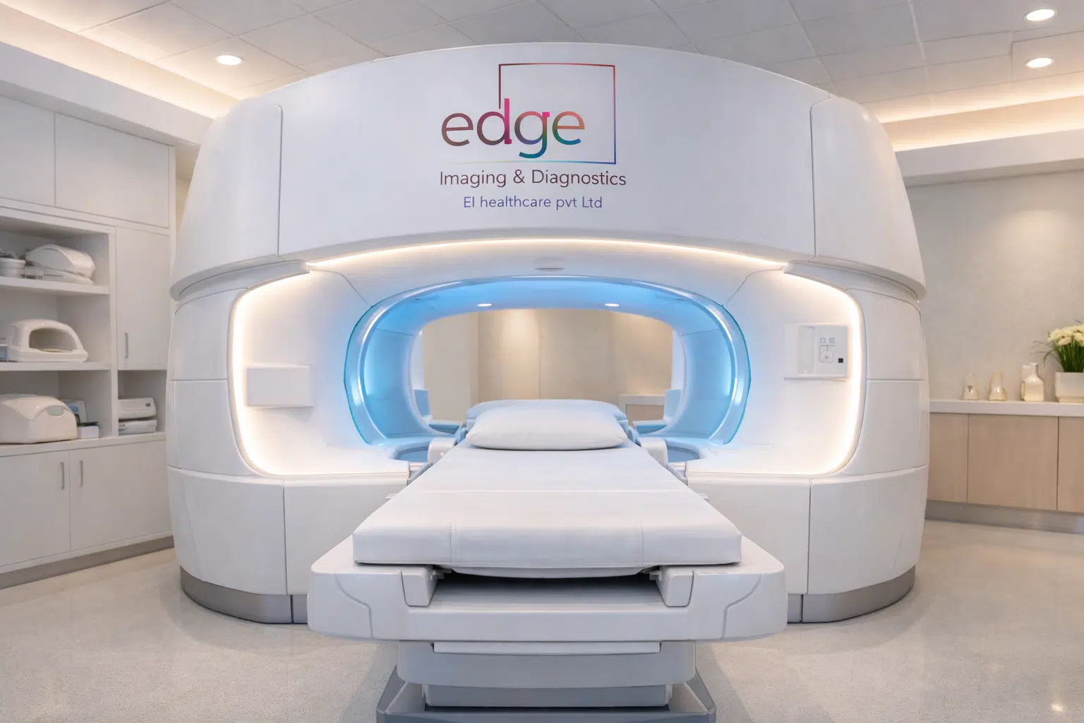
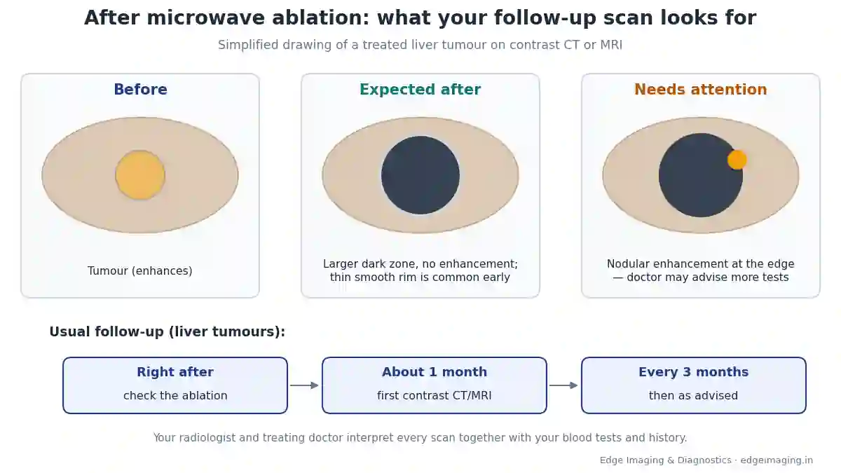
After microwave ablation, follow-up contrast CT or MRI scans check whether the tumour has been completely treated. A successful result usually shows a dark ablation zone, larger than the original tumour, with no enhancement inside. For liver tumours, scans are commonly done about one month after ablation and then every three months.
The ablation is over. You may have gone home the same day or the next morning. Now comes a new question that many patients and families find just as worrying: “How will we know it worked?”
The answer lies in your follow-up scans. This guide explains, in simple language, when these scans are done, what the radiologist looks for, and what the words on your report mean — from “ablation zone” and “non-enhancing” to “LR-TR nonviable”. It focuses mainly on liver tumours, where these terms are used most.
People search for this as “after microwave ablation”, “CT scan after microwave ablation”, “ablation zone meaning”, “how to know if ablation was successful” or “microwave ablation ke baad scan”. All are answered here, with a short Hinglish section at the end.
After microwave ablation: at a glance
- Why scans are needed: to confirm the tumour is fully treated and to catch any remaining or new disease early
- Usual timing (liver): a check right after the procedure, then about 1 month later, then every 3 months, as advised
- Scan type: contrast-enhanced CT or MRI
- Good sign: the treated area shows no enhancement inside
- Expected size: the ablation zone is usually larger than the original tumour, by design
- Common early finding: a thin, smooth rim around the zone, which usually fades within about a month
- Needs attention: thick, irregular or nodular enhancement at the edge of the zone
- Report language: LR-TR nonviable, LR-TR equivocal or LR-TR viable
What this guide covers
- A quick recap of microwave ablation
- Why follow-up scans matter
- The check right after the procedure
- The 1-month scan
- Every 3 months after that
- CT or MRI: which one?
- Why contrast is needed
- Preparing for your follow-up scan
- What the “ablation zone” is
- Why the zone looks bigger than the tumour
- “No enhancement”: the key good sign
- The thin rim around the zone
- Gas inside the zone early on
- How the zone changes over time
- Findings that need attention
- Residual tumour vs recurrence
- LR-TR categories, explained
- If your report says “equivocal”
- New spots elsewhere in the liver
- If some tumour is still active
- Kidney, lung and other organs
- Symptoms to report between scans
- Keep your scans together
- A sample report, read line by line
- CT scan after microwave ablation: what happens on the day
- “Arterial phase” and “washout”, explained
- Blood tests alongside your scans
- How long does follow-up continue?
- Living well between scans
- MRI after microwave ablation: what to expect
- More report words, explained simply
- For families supporting a patient
- Questions to ask your doctor
- Tips for scan day
- Coping with “scanxiety”
- Myths and facts
- Seedhi baat: microwave ablation ke baad scan
- Follow-up scans at Edge Imaging
- References
- Frequently asked questions
A quick recap of microwave ablation
Microwave ablation (MWA) is a minimally invasive treatment in which a thin needle-like probe is placed into a tumour under image guidance. Microwave energy heats and destroys the tumour tissue, along with a small margin of surrounding tissue. It avoids open surgery for many suitable patients.
If you want to understand the procedure itself, read our microwave ablation guide. This page is about what comes after: the scans that check the result.
Why follow-up scans matter
During ablation, the doctor aims to destroy the whole tumour. But a tumour cannot be checked by touch or by eye once it is treated — it stays inside the body. The only way to know whether every part was treated is imaging.
Follow-up scans do three jobs:
- Confirm complete treatment of the tumour that was ablated.
- Catch any remaining active tumour early, while further treatment is easier.
- Look for new tumours elsewhere, especially in the liver.
This is why keeping every follow-up appointment is as important as the ablation itself.
The check right after the procedure
A medical review on imaging after liver tumour treatment notes that the first post-treatment study is ideally performed immediately after treatment. This lets the treating doctor check, while you are still there, whether the ablation zone covers the whole tumour, and whether there is any immediate complication.
Your doctor will tell you what was seen. If more treatment was needed, it may have been done during the same session.
The 1-month scan
According to the same review, routine follow-up imaging is commonly performed about one month after ablation. By this time, much of the early swelling and inflammation around the treated area has settled, so the radiologist can judge the result more clearly.
This scan is often the most important one. It gives the first clear picture of whether the tumour has been completely treated.
Every 3 months after that
After the 1-month scan, follow-up is commonly done every three months for liver tumours, at least for a period of time. Your doctor may change this schedule depending on your type of tumour, your liver health, your blood tests and how things look on each scan.
Always follow the schedule your own doctor gives you — it is planned for your situation.
CT or MRI: which one?
Both contrast-enhanced CT and contrast-enhanced MRI are used to check the result of ablation. Your doctor chooses based on:
- What type of tumour was treated
- Which scan showed the tumour best before treatment
- Your kidney function and any allergies to contrast
- Whether you have metal implants or devices (relevant for MRI)
- Comparison with your previous scans
Using the same type of scan each time often makes comparison easier. If your doctor has asked for a particular scan, stick with it unless they advise a change.
Why contrast is needed
The key question after ablation is whether any part of the treated area still has a blood supply. Active tumour tissue usually has blood flowing to it; destroyed tissue does not.
A contrast agent injected into a vein travels with the blood. Areas with blood supply light up (“enhance”) on the scan. Areas without blood supply stay dark. That is why a plain scan without contrast usually cannot answer the question properly.
Preparing for your follow-up scan
- Bring all previous scans and reports — especially the scan done before ablation and the last follow-up. Comparison is the heart of the report.
- Bring your ablation summary or discharge papers.
- Kidney function test: your doctor may ask for a recent blood test before contrast.
- Fasting: follow the instructions given for your scan.
- Tell us about allergies, especially any previous reaction to contrast.
- For MRI: tell us about pacemakers, implants or metal in the body.
What the “ablation zone” is
The ablation zone is the area of tissue destroyed by the heat. It includes the tumour and a margin of surrounding tissue. On a contrast scan, it usually appears as a darker area where the original tumour used to be.
Your report may describe its size, shape and whether there is any enhancement in or around it. Those details are what tell your doctor whether the treatment worked.
Why the zone looks bigger than the tumour
Families are often surprised that the treated area looks bigger than the tumour did. This is expected, for two reasons:
- The margin is deliberate. The medical review notes that the treatment zone should be at least about 0.5–1.0 cm larger than the tumour in all directions, to make sure the edges are treated.
- Early swelling. Inflammation and swelling after treatment can make the zone look larger at first.
So a zone that looks larger than the original tumour is usually a sign that the treatment covered the tumour with a safety margin — not that something has grown.
“No enhancement”: the key good sign
This is the phrase most patients hope to see. A fully treated tumour should completely lack internal enhancement — meaning no blood supply is seen inside the treated area.
Your report may say “non-enhancing ablation zone”, “no residual enhancement” or “no evidence of viable tumour”. In simple words: on this scan, the treated area looks fully treated.
The thin rim around the zone
In the early weeks, the ablation zone may be surrounded by a thin, smooth, continuous rim of brightness. This is the body’s normal healing reaction to the heat. The medical review describes it as a normal post-ablation finding that usually disappears within about a month, though it may last longer.
What matters is the shape: a thin, smooth, even rim is expected. A thick, irregular or lumpy area is different, and is discussed below.
Gas inside the zone early on
Sometimes a scan soon after ablation shows small bubbles of gas inside the treated area. This can happen because of the heat. The review notes that gas early on should not necessarily be taken as a sign of infection. Your doctor will look at it together with how you feel, your temperature and blood tests.
How the zone changes over time
Over the following months, the ablation zone generally shrinks as the body slowly clears the destroyed tissue. On MRI, its appearance may also change. A zone that is getting smaller and stays non-enhancing is reassuring.
The zone may not disappear completely. A small scar-like area can remain for a long time; that alone is not a cause for concern.
Findings that need attention
The radiologist pays special attention to the edge of the ablation zone. According to the medical review:
- Any thickened or irregular enhancement at the margin should raise concern for active tumour.
- A typical warning sign is nodular enhancement along the edge in the arterial phase of the scan.
- Active tumour is almost always found at the periphery of the zone.
- Enhancement that grows or shows washout is more concerning than enhancement alone.
If your report mentions any of these, it does not automatically mean bad news, but your doctor will want to look closely and may advise further tests or treatment.
Residual tumour vs recurrence
Two words often confuse patients:
- Residual tumour: active tumour seen within about the first month. It usually means part of the tumour was not fully treated.
- Recurrence: active tumour seen later, after an earlier scan showed complete treatment.
The difference matters for planning, but in both cases the next step is a careful discussion with your treating team.
LR-TR categories, explained
For liver cancer (hepatocellular carcinoma), many radiologists use the LI-RADS Treatment Response system, written as LR-TR. It has three main categories:
| Category | What it means in simple words |
|---|---|
| LR-TR nonviable | No active tumour seen in the treated area — for example, no enhancement |
| LR-TR equivocal | Something is uncertain — for example, new or increasing enhancement without other typical features |
| LR-TR viable | Active tumour is likely present — for example, nodular or irregular enhancement at the edge |
| LR-TR nonevaluable | The scan could not be assessed properly, for example because of image quality or technique |
The American College of Radiology explains that these categories reflect the probability that tumour is still active after locoregional treatment, and that the system applies to treatments including microwave ablation, radiofrequency ablation and chemoembolization. “Nonviable” means no evidence of gross active tumour on the scan.
Your report may use these exact words. They help your doctors compare scans and make decisions in a consistent way.
If your report says “equivocal”
“Equivocal” means the radiologist cannot be sure yet. It is common to be asked to wait for the next scan, or to have a different type of scan, to clarify. Try not to panic: many equivocal findings turn out to be harmless healing changes. Ask your doctor what the plan is and when the next check will be.
New spots elsewhere in the liver
Follow-up scans look at the whole liver, not just the treated area. Sometimes a new spot appears in a different part of the liver. This is not a failure of the ablation — it is a separate finding. Finding it early on a routine scan is exactly why regular follow-up is advised.
If some tumour is still active
If a scan shows active tumour in or near the treated area, your treating team will discuss the options with you. Depending on the size, location and your overall health, these may include further treatment of the area or other approaches. Small residual areas found early are often easier to manage.
Keep all your scans and reports together so that the team can compare them quickly.
Kidney, lung and other organs
Microwave ablation is also used for some kidney, lung and other tumours. The basic idea of follow-up is similar — checking that the treated area has no active tumour — but the type of scan, the timing and the words used can differ. The LR-TR system described above is specific to liver cancer.
Your treating doctor will tell you which scans you need and when. Our guide to tumour ablation without surgery gives an overview of the organs treated.
Symptoms to report between scans
Do not wait for your next scheduled scan if you notice:
- Fever or chills
- Increasing pain in the treated area
- Yellowing of the eyes or skin
- Breathlessness or chest pain (especially after lung ablation)
- Blood in the urine (especially after kidney ablation)
- Feeling generally very unwell
Contact your treating doctor, or go to the nearest emergency department if symptoms are severe.
Keep your scans together
Comparison is the most valuable part of every follow-up report. Make a simple folder (paper or digital) with:
- The scan done before ablation
- The ablation procedure summary
- Every follow-up scan report and CD, in date order
- Blood test reports
Bring the whole folder to every scan and every doctor’s appointment.
A sample report, read line by line
Here is an example of report wording (not a real patient), with a simple explanation:
| Report line | What it means |
|---|---|
| “Post-ablation status of segment VI lesion” | The treated tumour was in segment VI of the liver |
| “Non-enhancing ablation zone measuring 3.8 × 3.4 cm” | The treated area is dark, with no blood supply, and larger than the original tumour by design |
| “Thin smooth peripheral rim enhancement, likely post-procedural” | An expected healing rim |
| “No nodular or irregular marginal enhancement” | No sign of active tumour at the edge |
| “No new focal lesion elsewhere in the liver” | No new spots seen |
| “Impression: LR-TR nonviable” | The treated area looks fully treated on this scan |
CT scan after microwave ablation: what happens on the day
A CT scan after microwave ablation is usually a multiphase contrast CT of the abdomen. In simple steps:
- Registration and safety check — allergies, kidney function and previous contrast reactions.
- A small cannula is placed in a vein in your arm for the contrast injection.
- You lie on the CT table and are asked to hold your breath for a few seconds at a time.
- Images are taken at different times after the contrast is injected, so the radiologist can see how the liver and the treated area take up the contrast.
- You can usually go home soon after, and drink water unless told otherwise.
The scan itself takes only a few minutes. The careful part is the reporting, when the radiologist compares the new images with your earlier scans.
“Arterial phase” and “washout”, explained
Your report may mention scan phases. After contrast is injected, it first reaches the arteries (the arterial phase), then the veins and the liver tissue (the portal venous phase), and later the delayed phase.
Many liver cancers take up contrast strongly in the arterial phase and then lose it faster than the surrounding liver in later phases — this is called washout. That is why radiologists look so carefully for any spot at the edge of the ablation zone that lights up early and then washes out. A treated area that stays dark in every phase is reassuring.
Blood tests alongside your scans
Your doctor will usually ask for blood tests along with your follow-up scans — for example, liver function tests and other tests relevant to your type of tumour and your liver health. Scans and blood tests are read together. A good scan with stable blood tests is the most reassuring combination. Bring the latest blood reports to your scan and your appointment.
How long does follow-up continue?
There is no single answer for everyone. For liver tumours, follow-up is commonly every three months at first. Your doctor may adjust the schedule over time depending on your scans, your blood tests, the type of tumour and the health of your liver. Some patients also need regular checks because their liver condition itself needs monitoring. Ask your doctor to explain your personal schedule and write it down.
Living well between scans
- Take your medicines exactly as prescribed, including treatment for any underlying liver disease.
- Avoid alcohol if your doctor advises it — it protects the liver.
- Eat a balanced diet and stay as active as your doctor allows.
- Keep a symptom diary — note any pain, fever or yellowing to discuss with your doctor.
- Book your next scan early, so it is never delayed.
MRI after microwave ablation: what to expect
If your doctor prefers MRI, the scan is usually done with a contrast injection and takes longer than CT — often 30 to 45 minutes or more, depending on the protocol. You lie still inside the scanner, wear ear protection because MRI is noisy, and follow breathing instructions. MRI uses no X-ray radiation. Tell us in advance about any pacemaker, implant or metal in your body, and if you feel anxious in closed spaces.
On MRI, the treated area may change in appearance over the months, as the destroyed tissue is slowly cleared. The radiologist compares each scan with the previous one to make sure the changes are the expected healing pattern.
More report words, explained simply
| Word | Simple meaning |
|---|---|
| Post-ablation changes | Expected changes after treatment |
| Perilesional | Around the treated area |
| Marginal / peripheral | At the edge of the treated area |
| Hyperenhancement | Brighter after contrast than the surroundings |
| Stable | No change since the last scan |
| Interval decrease | Smaller than on the last scan |
| Cirrhotic liver | A scarred liver, a common background in liver cancer |
| No new lesion | No new spot seen anywhere else |
For families supporting a patient
Family members can make a big difference: keep a calendar of scan dates and blood tests, maintain the folder of reports, accompany the patient on scan days, and note questions for the doctor. Most of all, be patient with the worry that often comes before each scan. A calm, organised routine helps everyone through months of follow-up.
Questions to ask your doctor
- Does the report show the tumour is fully treated?
- Is the rim around the zone expected at this stage?
- When is my next scan, and should it be CT or MRI?
- Do I need blood tests before or alongside the scan?
- If the report is equivocal, what is the plan?
- Is there anything new elsewhere in the liver?
- Which symptoms should make me call you before the next scan?
Tips for scan day
- Book follow-up scans well in advance so they are on time.
- Carry your full folder of previous scans.
- Drink water after a contrast scan, unless told otherwise.
- Ask when the report will be ready, and plan your doctor’s appointment after it.
- Bring a family member for support if you feel anxious.
Coping with “scanxiety”
Many patients feel anxious in the days before each scan and while waiting for the report. This is so common that it has a nickname: “scanxiety”. It can help to plan something calming on scan day, to ask for the report time in advance, and to arrange the doctor’s appointment soon after so you are not left waiting. Remember that regular follow-up is your safety net — it exists to catch problems early.
Myths and facts
| Myth | Fact |
|---|---|
| “The treated area looks bigger, so the tumour has grown.” | The zone is meant to be larger, to include a safety margin; early swelling adds to it. |
| “Any rim on the scan means cancer is back.” | A thin, smooth rim is a common healing finding early on. |
| “If the 1-month scan is clear, no more scans are needed.” | Regular follow-up continues, commonly every 3 months for liver tumours, as advised. |
| “A plain scan is enough to check.” | Contrast is usually needed to see whether any blood supply remains. |
| “A new spot elsewhere means the ablation failed.” | New spots are separate findings; finding them early is the purpose of follow-up. |
Seedhi baat: microwave ablation ke baad scan
Microwave ablation ke baad scan kyun? Yeh dekhne ke liye ki tumour poori tarah treat hua ya nahi, aur koi naya spot to nahi bana.
Kab-kab scan hota hai? Liver tumour me aam taur par procedure ke turant baad check, phir lagbhag 1 mahine baad contrast CT ya MRI, aur phir har 3 mahine — jaise doctor kahein.
Report me achha sign kya hai? “No enhancement” ya “non-enhancing ablation zone” — matlab treated jagah me khoon ka flow nahi dikh raha, tumour treat ho chuka lag raha hai.
Treated jagah badi kyun dikhti hai? Kyunki doctor jaan-boojh kar tumour ke chaaron taraf thoda extra hissa bhi treat karte hain, aur shuru me thodi soojan bhi hoti hai.
LR-TR ka matlab? Nonviable = tumour active nahi dikh raha. Equivocal = abhi pakka nahi, agla scan dekhna hoga. Viable = kuch active tumour ho sakta hai, doctor aage ka plan batayenge.
Follow-up scans at Edge Imaging
At Edge Imaging & Diagnostics, West Delhi, we perform contrast-enhanced CT and MRI scans for follow-up after tumour ablation, with careful comparison to your previous scans. Bring your earlier reports and CDs, and your scan will be reported with that comparison in mind. Read more about radiofrequency ablation and microwave ablation for tumour patients.
Send your doctor’s prescription on WhatsApp, and we will share the earliest slot, how to prepare for contrast, and today’s actual pricing.
💬 WhatsApp to book your follow-up scan | Call 9555999960
References
- MRI assessment of hepatocellular carcinoma after local-regional therapy: a comprehensive review — Mendiratta-Lala M, et al. Radiology: Imaging Cancer, 2020.
- What are LI-RADS treatment response categories? — American College of Radiology (ACR) RADS support.
Frequently asked questions
When is the first scan after microwave ablation?
A check is often done right after the procedure. For liver tumours, routine follow-up contrast CT or MRI is commonly done about one month after ablation.
How often are scans needed after ablation?
For liver tumours, follow-up is commonly every three months after the first month, for a period advised by your doctor based on your condition.
What does “ablation zone” mean?
It is the area of tissue destroyed by the heat, including the tumour and a safety margin. It usually appears darker than the surrounding liver on a contrast scan.
Why is the ablation zone bigger than my tumour?
The treatment aims to cover the tumour plus a margin of about 0.5 to 1 cm all around, and early swelling can make the zone look larger.
What does “no enhancement” mean on my report?
It means no blood supply is seen inside the treated area, which is the key sign that the tumour has been fully treated on that scan.
Is a rim around the ablation zone a bad sign?
A thin, smooth, even rim is a common healing finding early on and usually fades within about a month. Thick, irregular or nodular enhancement needs attention.
What findings suggest the tumour is still active?
Thickened, irregular or nodular enhancement at the edge of the zone, especially if it grows or shows washout, raises concern for active tumour.
What is the difference between residual tumour and recurrence?
Residual tumour is active tumour seen within about the first month. Recurrence is active tumour seen later, after a scan had shown complete treatment.
What does LR-TR nonviable mean?
It means no active tumour is seen in the treated area on that scan, using the LI-RADS treatment response system for liver cancer.
What does LR-TR equivocal mean?
It means the findings are uncertain. Your doctor may advise waiting for the next scan or doing another type of scan to clarify.
What does LR-TR viable mean?
It means active tumour is likely present in or near the treated area. Your treating team will discuss further options with you.
Why is contrast needed for follow-up scans?
Contrast shows whether any part of the treated area still has a blood supply, which is how active tumour is detected.
Is gas in the ablation zone a sign of infection?
Not necessarily. Gas can appear early because of the heat. Your doctor will judge it along with your symptoms and blood tests.
Microwave ablation ke baad scan kab hota hai?
Liver tumour me aam taur par turant baad check, lagbhag 1 mahine baad contrast CT ya MRI, aur phir har 3 mahine, jaise doctor kahein.
Report me “no enhancement” ka matlab kya hai?
Matlab treated jagah me khoon ka flow nahi dikh raha, yaani us scan par tumour poori tarah treat hua lag raha hai.
Do kidney and lung ablations follow the same schedule?
The idea is similar, but the scan type, timing and terms can differ. LR-TR is specific to liver cancer. Follow your doctor’s schedule.
Where can I get follow-up CT or MRI after ablation in West Delhi?
At Edge Imaging & Diagnostics, West Delhi. WhatsApp or call 9555999960 with your prescription for the earliest slot and today’s actual pricing.
The bottom line
After microwave ablation, follow-up scans are how you and your doctor know the treatment worked. A dark, non-enhancing ablation zone — often larger than the original tumour, sometimes with a thin healing rim early on — is what doctors hope to see. Irregular or nodular enhancement at the edge needs a closer look.
Keep every appointment, bring all your previous scans, and ask your doctor to walk you through each report. Regular follow-up is your best protection.
Many patients go through months of follow-up with reports that stay reassuring scan after scan. If something does need attention, finding it early on a planned scan gives you and your doctors the most options. That is the real value of follow-up imaging after microwave ablation: not worry, but early, clear information that keeps you one step ahead.
If you are unsure about any word on your report, write it down and ask. No question is too small when it comes to your health.
Written by the medical team at Edge Imaging & Diagnostics, West Delhi. Medically reviewed by Dr. Nishant Thapar. Last reviewed: October 2026.
Disclaimer: this article is general information to help patients understand follow-up scans after ablation. It is not a diagnosis. The details above refer mainly to liver tumours; follow-up for other organs may differ. Always follow your treating doctor’s advice.