
 PV
 TG
 MN
 EM
 EI
 ND

 CT Scan
 PET CT
 Digital X-ray
 Ultrasound
 Amniocentesis

 Fetal Reduction
 Thermalytix Breast Screening
 Cardiology
 Lab Tests
 GAMCA Pre-Medical

An AV malformation (arteriovenous malformation, or AVM) is a tangle of abnormal blood vessels in which arteries connect directly to veins, without the normal tiny capillaries in between. In the arms, legs and body, an AVM can cause a warm, pulsating swelling, pain, skin darkening, wounds or bleeding. The main treatment for most body and limb AVMs is image-guided embolization: closing the abnormal tangle from inside the blood vessels, often over more than one session, sometimes combined with surgery.
Many patients first notice a soft swelling on the hand, foot, leg, arm, buttock or chest wall that feels warm and seems to throb. Some are told it is a “birthmark”, a “vein problem” or a “lump”. Others live with it for years until it grows after puberty, pregnancy or an injury, or starts to hurt, bleed or form a wound.
This guide explains AVMs of the body and limbs in simple language: what they are, how they differ from other vascular malformations and varicose veins, the warning signs, which scans are needed, how treatment works, what recovery looks like, and the honest results and risks. It is written for patients and families, not doctors.
People search for this as “AV malformation treatment”, “AVM treatment”, “arteriovenous malformation treatment”, “AVM in leg”, “AVM in hand”, “peripheral AVM”, “AVM embolization”, “AVM treatment in Delhi” or, in Hinglish, “AVM kya hota hai aur iska ilaj”. All of these are answered here, with a short Hinglish section near the end.
AV malformation treatment (body and limbs): at a glance
- What it is: a fast-flow tangle of vessels where arteries join veins directly
- Usually present from birth, but may grow with puberty, pregnancy, injury or surgery
- Common signs: warm, pulsating swelling, pain, skin changes, wounds or bleeding
- First test: colour Doppler ultrasound
- Planning scans: CT angiography, MR angiography and, at treatment, catheter angiography
- Main treatment: image-guided embolization of the abnormal tangle (the nidus)
- Other options: sclerotherapy in selected cases, surgery for well-defined AVMs, compression and pain relief
- Sessions: often more than one; long-term follow-up is needed
- Brain and spine AVMs: a different condition, treated by neurovascular teams
What this guide covers
- What an AV malformation is
- AVMs and other vascular malformations
- Where AVMs occur in the body and limbs
- What causes an AVM?
- Why AVMs can grow over time
- Symptoms of an AVM in the arm, leg or body
- The four stages of an AVM (Schobinger stages)
- AVM vs varicose veins vs venous malformation
- When to seek urgent care
- How an AVM is diagnosed
- Doppler ultrasound: the first scan
- CT angiography and MR angiography: mapping the AVM
- Does every AVM need treatment?
- The goal of AVM treatment
- Embolization: the main treatment
- Where sclerotherapy fits in
- Surgery for AVMs
- Compression, pain relief and watchful waiting
- Why simply tying off the artery does not work
- Before AVM treatment
- The embolization procedure, step by step
- After the procedure: recovery
- How many sessions are needed?
- Results: what to expect honestly
- Risks and complications
- Warning signs after treatment
- Follow-up and recurrence
- AVMs, pregnancy and hormones
- AVMs in children and teenagers
- Living with an AVM
- What about brain and spinal AVMs?
- What affects the cost of AVM treatment
- Questions to ask your doctor
- Seedhi baat: AVM kya hai aur iska ilaj
- AVM diagnosis and treatment at Edge Imaging, West Delhi
- References
- Frequently asked questions
What an AV malformation is
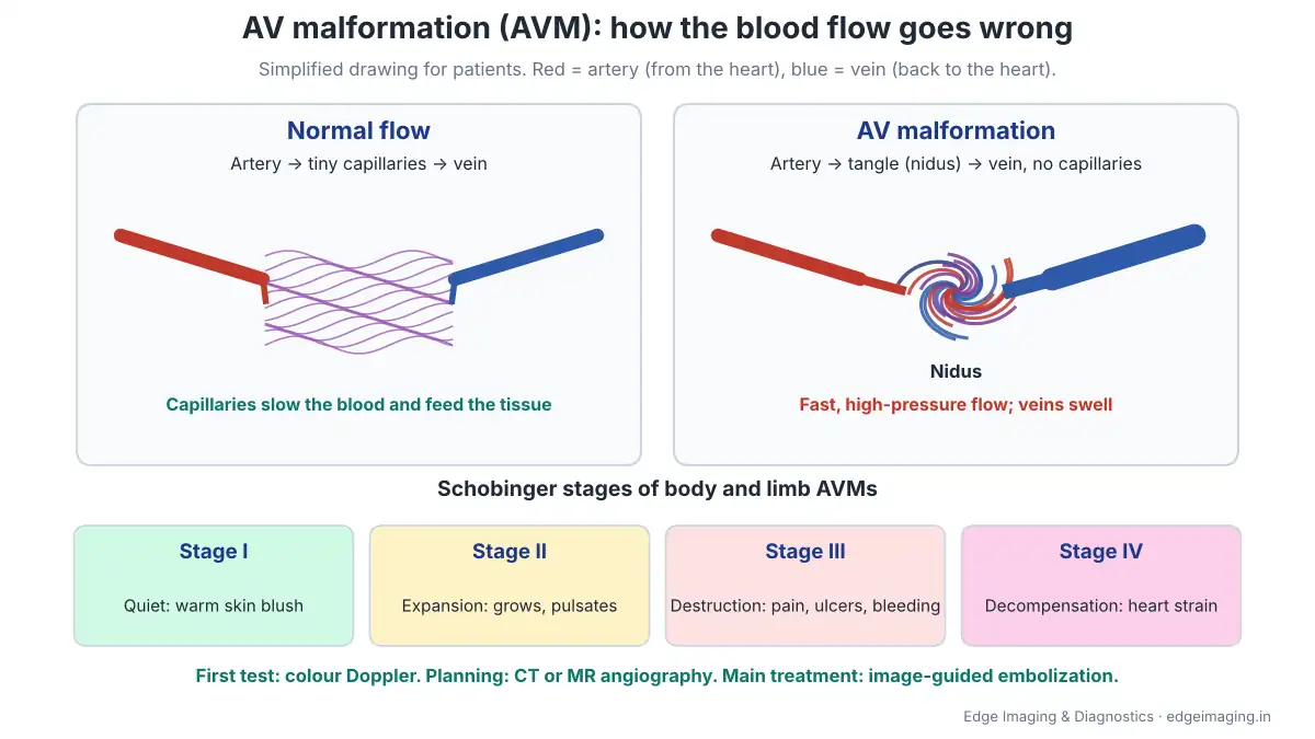
Normally, blood leaves the heart in arteries, passes through millions of tiny vessels called capillaries that feed the tissues, and returns to the heart in veins. Capillaries act like a gentle bridge that slows the blood down.
In an arteriovenous malformation (AVM), that bridge is missing. Arteries connect directly to veins through a tangle of abnormal vessels. Doctors call this tangle the nidus, from the Latin word for nest. Cleveland Clinic describes an AVM as an abnormal tangle of blood vessels that looks like a bird’s nest.
Because there are no capillaries to slow it down, blood rushes at high pressure from the arteries into the veins. Over time this can:
- Stretch and enlarge the veins around the AVM
- “Steal” blood from nearby tissues, which may then get too little oxygen
- Cause pain, skin damage, wounds and bleeding
- In large AVMs, make the heart work much harder
AVMs are not cancer. They are a problem in how blood vessels formed, and they are uncommon.
AVMs and other vascular malformations
The International Society for the Study of Vascular Anomalies (ISSVA) groups vascular anomalies into two families: vascular tumours (such as infantile haemangioma) and vascular malformations. Malformations are further divided by how fast blood flows through them:
- Slow-flow malformations: capillary malformations (such as port-wine stains), venous malformations and lymphatic malformations
- Fast-flow malformations: arteriovenous malformations (AVMs) and arteriovenous fistulas (AVFs)
Johns Hopkins Medicine notes that low-flow malformations are more common than high-flow ones, and a 2024 study from a German vascular anomaly centre states that AVMs account for less than 3% of vascular malformations. This matters because the type decides the treatment. A slow-flow venous malformation is often treated very differently from a fast-flow AVM.
An arteriovenous fistula is a single direct connection between an artery and a vein, sometimes after an injury, while an AVM has many small connections inside a nidus.
Where AVMs occur in the body and limbs
AVMs can occur almost anywhere. Outside the brain and spine, they are often called peripheral or extracranial AVMs. Common places include:
- Hands and fingers
- Feet and toes
- Legs: thigh, knee, calf, ankle
- Arms: upper arm, forearm, elbow
- Buttocks and pelvis
- Chest wall, back and abdominal wall
- Internal organs such as the lungs, liver, kidneys or uterus
This guide focuses mainly on AVMs of the limbs and the trunk (chest wall, back, abdominal wall and buttocks). AVMs inside organs and in the brain and spine need organ-specific or neurovascular planning; the brain and spine are covered briefly later.
What causes an AVM?
Most AVMs are believed to be present from birth, forming while the baby’s blood vessels develop in the womb. Cleveland Clinic notes that scientists are not sure exactly what causes them. Certain inherited conditions, such as hereditary haemorrhagic telangiectasia, raise the chance of AVMs, but most AVMs are not inherited.
Nothing the parents did in pregnancy causes an AVM, and it is not caused by diet, exercise or lifestyle.
Why AVMs can grow over time
An AVM may look like a small birthmark or may not be visible at all in childhood. A 2024 study from a German vascular anomaly centre describes AVMs as present at birth but growing and likely progressing with age, and notes that puberty, trauma and pregnancy may be associated with a risk of worsening. The UK’s Butterfly AVM Charity also lists puberty, pregnancy, injury and surgery as possible triggers.
Progression is very variable. Some AVMs stay quiet for decades; others grow over a few years. This is why a doctor experienced in vascular malformations should guide when to treat and when to watch.
Symptoms of an AVM in the arm, leg or body
Symptoms depend on the size and location of the AVM and how much blood flows through it. Common signs include:
- A swelling or lump that may slowly enlarge
- Warmth over the area, compared with the other side
- A pulsation you can see or feel, or a buzzing feeling (thrill)
- A pink, red or bluish skin stain over the area
- Enlarged, bulging veins around it
- Pain, especially with activity or when the limb hangs down
- Dark, thickened or shiny skin, or a wound that does not heal
- Bleeding from the skin, sometimes heavy
- Overgrowth of one limb, which may be longer or larger than the other
- In very large AVMs: breathlessness, tiredness and leg swelling from strain on the heart
In the 2024 German study of 60 patients with AVMs outside the brain, the most common symptom was pain (81%), followed by pulsation (64%) and local warmth (62%).
The four stages of an AVM (Schobinger stages)
Doctors describe how active a body or limb AVM is using the Schobinger staging:
- Stage I, quiescence: a skin blush or stain, warmth, and fast-flow shunting seen on Doppler ultrasound
- Stage II, expansion: the AVM enlarges, with pulsation, a thrill you can feel and a bruit (whooshing sound) a doctor can hear
- Stage III, destruction: pain, skin changes, ulcers and bleeding
- Stage IV, decompensation: heart failure from the strain of the shunt
The stage helps decide whether to treat and how urgently. In the 2024 German study, most patients (57%) were in stage II when first assessed. Stage III and IV AVMs need prompt specialist assessment.
AVM vs varicose veins vs venous malformation
Because an AVM often shows enlarged veins on the surface, it is sometimes mistaken for varicose veins. The differences matter:
- Varicose veins: leaky valves in normal leg veins cause slow pooling of blood. No pulsation, no warmth. Treated with methods such as foam sclerotherapy or vein ablation.
- Venous malformation: a slow-flow, spongy, bluish swelling present from birth, which often swells when the limb hangs down. Usually not warm or pulsating. Often treated with sclerotherapy.
- AVM: fast flow, often warm and pulsating, with a buzz or whooshing sound. Needs careful planning, usually with embolization.
Treating an AVM as if it were simple varicose veins, for example by stripping or injecting only the visible veins, can make it worse. A Doppler scan quickly shows whether the flow is slow or fast. If you have only small thread-like veins, see our guide to spider veins treatment.
When to seek urgent care
Go to an emergency department immediately if:
- An AVM is bleeding heavily and does not stop with firm pressure
- A finger, toe or part of a limb becomes cold, pale, blue or very painful
- You have sudden breathlessness, chest pain or fainting
- A wound over the AVM becomes red, hot, swollen and painful with fever
For bleeding, press firmly on the spot with a clean cloth, raise the limb if possible, and get help.
How an AVM is diagnosed
Diagnosis starts with a careful history and examination. The doctor looks at the swelling, feels for warmth and pulsation, and listens for a bruit. Then imaging confirms the diagnosis and shows the AVM’s size and blood supply:
- Colour Doppler ultrasound: the first test, confirming fast flow
- CT angiography (CTA): a contrast CT scan that maps arteries, the nidus and veins in 3D
- MRI and MR angiography (MRA): show the AVM’s extent in muscles and soft tissues, without radiation
- Catheter angiography (DSA): usually done as part of treatment, to see the nidus in real time
Johns Hopkins Medicine notes that deeper vascular malformations can be diagnosed using advanced imaging, preferably MRI. The 2024 German study found that ultrasound is useful for initial diagnosis but not enough for treatment planning, and that MR angiography, dynamic CT angiography and angiography are important planning tools.
Doppler ultrasound: the first scan
A colour Doppler ultrasound is painless and uses no radiation. Gel is applied, and a probe is moved over the swelling. The machine shows blood vessels in colour and measures how fast blood flows.
In an AVM, Doppler typically shows:
- Many tangled vessels with very fast, turbulent flow
- Arteries feeding the area with abnormal, low-resistance flow
- Veins draining it with a pulsating, “arterialised” pattern
These findings separate an AVM from a slow-flow venous malformation, a cyst or a solid lump. For the limbs, a scan of the arteries such as an arterial Doppler can also check blood supply to the hand or foot beyond the AVM.
CT angiography and MR angiography: mapping the AVM
Before treatment, the doctor needs a detailed map: which arteries feed the AVM, where the nidus is, how big it is, which veins drain it, and which normal structures lie close by.
CT angiography is fast and shows the arteries and veins in sharp 3D detail. A contrast injection is given through a vein in the arm, and the scan takes only a few seconds. For the legs, this is the same type of scan as a CT peripheral angiography. You will be asked about kidney function, diabetes medicines and allergies before contrast.
MRI and MR angiography show how far the AVM spreads into muscles, fat and bone. Because MRI uses no radiation, it is useful in children and for repeat scans.
Your doctor chooses the right combination of scans for your AVM.
Does every AVM need treatment?
Not always. Treatment is considered when the benefits are likely to be greater than the risks. Common reasons to treat include:
- Pain that affects daily life or work
- Bleeding, or a skin wound that does not heal
- Steady growth of the AVM
- Problems using the hand, foot or limb
- Signs of strain on the heart
- A disfiguring swelling causing serious distress
A small, quiet stage I AVM with no symptoms may simply be watched with regular check-ups and scans. Incomplete or poorly planned treatment can sometimes stimulate an AVM to grow, so “doing something” is not always better than careful watching.
The goal of AVM treatment
The 2024 German study states that occlusion of the nidus is the mainstay of treatment. In other words, the aim is to close the abnormal tangle itself, not just the arteries feeding it.
Realistic goals are:
- Reduce or stop pain
- Stop bleeding and help wounds heal
- Shrink the swelling and slow or stop growth
- Protect the hand, foot or limb function
- Reduce strain on the heart in large AVMs
Complete cure is possible in some small, well-defined AVMs. For larger or diffuse AVMs, the honest goal is often good long-term control with fewer symptoms.
Embolization: the main treatment
Embolization means blocking abnormal blood vessels from inside, using a material delivered through a needle or a fine tube (catheter), under X-ray guidance. Johns Hopkins Medicine describes embolization as very effective in managing AVMs and typically the first-line treatment.
The material is placed into the nidus, where it closes the abnormal connections. Depending on the AVM, it can be delivered:
- Through the artery (transarterial): a catheter is guided from an artery, usually in the groin or wrist, to the AVM
- By direct puncture: a needle is passed through the skin straight into the nidus, guided by ultrasound or X-ray
- Through the vein (transvenous): in some AVMs, the draining vein is reached and closed close to the nidus
Materials used around the world include liquid embolic agents (such as special glues and polymers), absolute alcohol and coils. The Butterfly AVM Charity describes embolization as a main treatment, usually done in several sessions over time, with the aim of closing the shunts themselves using permanent agents. The choice of route and material depends on the AVM’s size, location, flow pattern and nearby nerves and skin, and is made by the treating interventional radiologist after imaging.
Where sclerotherapy fits in
Sclerotherapy means injecting a medicine that irritates and closes abnormal vessels. It is the main treatment for slow-flow venous and lymphatic malformations.
For fast-flow AVMs, some sclerosing agents are used together with embolization in selected cases, especially in the draining part of the AVM. For example, a 2025 study of patients with large limb AVMs causing heart failure used coils and sclerosing agents, with an average of two sessions per patient, and reported major improvement in heart symptoms.
So if you have read that “AVM is treated with injections”, it is partly true: the injections are given into the AVM under imaging guidance as part of a planned embolization and sclerotherapy treatment, not as a simple clinic injection into a visible vein.
Surgery for AVMs
Surgery to remove an AVM may be considered when it is small, localised and well-defined, for example in a finger or a small area of the skin and muscle. The 2024 German study notes that resection, with or without embolization beforehand, has shown a lower recurrence rate in earlier research, and that embolization can be done before surgery to reduce blood loss.
For large or diffuse AVMs, complete surgical removal is often difficult. Johns Hopkins Medicine notes that complete removal is often difficult and that high-flow malformations can be challenging to treat with surgery. A team approach between interventional radiology and surgery gives the best chance in such cases.
Compression, pain relief and watchful waiting
Not every patient needs a procedure straight away. Supportive care can help:
- Compression garments for an arm or leg can reduce swelling and aching
- Pain relief as advised by your doctor
- Skin care to protect fragile skin, and prompt care of any wound
- Avoiding injury to the area, especially in contact sports
- Regular review with Doppler or MRI to watch for growth
Research into medicines that act on the genetic pathways behind AVMs is ongoing worldwide, but these are not yet standard treatment for most patients.
Why simply tying off the artery does not work
It may seem logical to cut off the AVM’s blood supply by tying or blocking the main artery feeding it. In practice, this usually fails and can make future treatment harder. Johns Hopkins Medicine explains that the nidus can form new arterial connections, letting the AVM regrow. Blocking the feeding artery also removes the doctor’s route to reach the nidus later.
The Butterfly AVM Charity similarly notes that coils may close large shunts but should not block the feeding arteries. This is why AVM treatment should be planned by doctors with specific experience in vascular malformations.
Before AVM treatment
- Bring all scans: Doppler reports, CT or MR angiography, and any earlier treatment records
- Blood tests: blood count, kidney function and clotting tests are commonly checked
- Medicines: tell the doctor about blood thinners, diabetes medicines (especially metformin before contrast), and allergies. Do not stop any medicine on your own.
- Fasting: you will be told how long to fast if sedation or anaesthesia is planned
- Consent: the doctor explains the plan, expected benefits, risks, and the likely number of sessions
- Pregnancy: tell the team if you are or might be pregnant
The embolization procedure, step by step
- Preparation: you change into a gown, an IV line is placed, and monitoring is started.
- Anaesthesia: local anaesthesia with sedation, or general anaesthesia, depending on the AVM and the materials used.
- Access: a small needle puncture is made in an artery (often in the groin or wrist), or directly into the AVM, guided by ultrasound.
- Angiography: contrast is injected to map the nidus on real-time X-ray.
- Embolization: the embolic material is carefully delivered into the nidus, in small amounts, while the doctor watches the flow.
- Check: repeat images confirm how much of the AVM has been closed.
- Closure: the catheter is removed and pressure is applied to the puncture site.
Approaches vary between centres and between patients. The procedure time varies with the size and complexity of the AVM. Depending on the procedure, you may go home the same day or be kept under observation for longer; your doctor will tell you in advance.
After the procedure: recovery
- Swelling and pain in the treated area are common for several days and are managed with medicines and elevation
- Bruising at the puncture site is common
- Rest and elevation of the limb help the swelling settle
- Wound care: keep the puncture site clean and dry as instructed
- Activity: gentle walking is usually encouraged; avoid heavy lifting or strenuous exercise for the period your doctor advises
- Follow-up visit: to check healing and plan the next step
The treated area may feel firm for weeks as the closed vessels are gradually absorbed and replaced by scar tissue.
How many sessions are needed?
Many body and limb AVMs need more than one session. Johns Hopkins Medicine notes that AVMs often require a series of treatments to achieve complete or near-complete resolution. In the 2025 study of large limb AVMs mentioned above, patients had an average of two sessions, with a range of one to three.
Treating in stages lets the tissues recover between sessions and allows the doctor to reassess the AVM before the next step.
Results: what to expect honestly
Results vary widely with the size, location and stage of the AVM:
- Small, well-defined AVMs may be closed completely, sometimes with surgery added
- Many patients have clear improvement in pain, bleeding and swelling
- Large, diffuse AVMs are often controlled rather than cured
- Long-term follow-up is needed because AVMs can regrow
Studies report mixed outcomes. In the 2024 German series, 25% of patients were free of symptoms at last contact, while about one-third still had symptoms or progression and many were lost to follow-up. In the 2025 study of very large limb AVMs with heart failure, most patients achieved 80% or more closure of the malformation and major improvement in breathlessness. These are specialist-centre results and differ from patient to patient.
The key message: AVM treatment is a long-term partnership, not a one-time fix. Steady, careful treatment usually gives better results than one aggressive attempt.
Risks and complications
Every AVM treatment carries risks, which depend on the location, the material used and how much of the AVM is treated. They include:
- Pain and swelling after treatment, usually temporary
- Skin injury: blistering, skin breakdown or a wound over the treated area
- Nerve irritation or injury: numbness, tingling or weakness, usually temporary but occasionally lasting
- Non-target embolization: material reaching normal vessels, which can reduce blood supply to nearby tissue
- Delayed wound healing
- Bleeding or bruising at the puncture site
- Contrast reactions or effects on kidney function
- Blood clots in veins
In the 2024 German series, the overall complication rate was 39%, mostly delayed wound healing and nerve irritation, and the authors note that rates vary with the embolic agent, AVM location and extent. Ask your doctor what the specific risks are for your AVM.
Warning signs after treatment
Contact your doctor urgently if you notice:
- Increasing pain that is not controlled by the prescribed medicines
- A finger, toe or part of the limb becoming cold, pale, blue or numb
- Blistering, blackening or breakdown of the skin
- Bleeding or a growing swelling at the puncture site
- Fever, redness spreading from the area, or pus
- Breathlessness or chest pain
Follow-up and recurrence
After treatment, follow-up usually includes clinic visits and repeat imaging, such as Doppler ultrasound or MRI, to check how much of the AVM remains and whether it is regrowing. For AVMs in general, Cleveland Clinic suggests follow-up visits every three months in the first year, then yearly; your doctor will set a schedule for your AVM.
AVMs can come back, especially if part of the nidus remains. Growth is more likely around puberty, pregnancy or after injury. If symptoms return, further treatment can usually be planned.
AVMs, pregnancy and hormones
Pregnancy may be associated with growth or worsening of an AVM, and Cleveland Clinic notes that pregnancy can increase bleeding risk in AVMs. If you have an AVM and are planning a pregnancy, discuss it with your treating doctor and obstetrician in advance.
Procedures that use X-rays and contrast are usually avoided during pregnancy unless there is an emergency. Doppler ultrasound and, where needed, MRI without contrast can be used for monitoring.
AVMs in children and teenagers
Many AVMs are first noticed in childhood as a warm birthmark or a swelling. Not every child needs early treatment; many are watched carefully, because the risks of treatment must be balanced against a growing body.
Teenage years and puberty are a common time for AVMs to grow. Parents should watch for increasing size, warmth, pain, skin changes or bleeding, and keep regular follow-up with a doctor experienced in vascular malformations.
Living with an AVM
- Keep copies of all scans and treatment records, and carry a summary when travelling
- Protect the area from injury; choose sports and work with care
- Use compression garments if advised
- Know how to press firmly on a bleeding spot and when to seek help
- Look after your general heart health: blood pressure, weight and exercise
- Do not ignore emotional strain; living with a visible or painful condition is hard, and support helps
What about brain and spinal AVMs?
AVMs in the brain and spinal cord are a different condition with different risks, mainly bleeding inside the head or spine. Cleveland Clinic gives the yearly chance of bleeding from an unruptured brain AVM as about 2 to 4%. They are treated by specialised neurovascular teams with neurosurgery, neuro-interventional embolization or radiosurgery.
If you have symptoms such as a sudden severe headache, seizures, weakness or speech problems, seek emergency care. For known or suspected brain AVMs, imaging such as MR angiography of the brain or CT brain angiography helps your neurologist or neurosurgeon plan care.
What affects the cost of AVM treatment
AVM treatment is highly individual, so there is no single fixed price. The cost usually depends on:
- The size, location and stage of the AVM
- The scans needed: Doppler, CT angiography, MRI
- The type and amount of embolic material used
- Whether sedation or general anaesthesia is needed
- Observation time after the procedure, if needed, and medicines
- The number of sessions
- Follow-up imaging
The best way to know the actual cost for your AVM is to share your reports and get a personalised plan.
Questions to ask your doctor
- Is this an AVM, a fistula, or a slow-flow malformation?
- What stage is my AVM, and does it need treatment now?
- What scans do I need before treatment?
- Which route and material do you plan to use, and why?
- How many sessions are likely?
- What are the specific risks for my AVM’s location?
- Will surgery be needed as well?
- How will we follow up, and how often?
Seedhi baat: AVM kya hai aur iska ilaj
AVM yaani arteriovenous malformation me khoon ki nasein janam se hi galat tarike se judi hoti hain. Artery seedhe vein se jud jaati hai, beech ki patli nasein (capillaries) nahi hoti. Isse haath, pair ya sharir ke kisi hisse me garam, dhadakti hui sujan, dard, kaali skin, ghaav ya khoon nikalna ho sakta hai.
Pehle colour Doppler se jaanch hoti hai, phir CT angiography ya MRI se poora naksha banta hai. Mukhya ilaj embolization hai, jisme nas ke andar se ya seedhe sui se AVM ke guchhe (nidus) ko band kiya jaata hai. Aksar ek se zyada session lagte hain aur baad me regular follow-up zaroori hai.
Sirf upar wali nas ko baandh dena ya varicose veins samajh kar ilaj karna AVM ko bigaad sakta hai. Isliye anubhavi doctor se hi plan karwayein. Agar khoon tez beh raha ho, ya ungli/pair thanda aur neela pad jaye, toh turant emergency jaayein.
AVM diagnosis and treatment at Edge Imaging, West Delhi
At Edge Imaging & Diagnostics, West Delhi, we diagnose AVMs with colour Doppler ultrasound and CT angiography, and we provide image-guided, minimally invasive treatment for AVMs of the body and limbs, such as the arms, legs, hands, feet and trunk wall. The treatment plan is made after imaging, and the number of sessions is discussed with you in advance.
We do not treat AVMs of the brain or spinal cord. For these, we can help with imaging, and we will guide you towards the right neurovascular team.
Send your Doppler, CT or MRI reports, or clear photos of the swelling, on WhatsApp. We will tell you the next step, the earliest slot, what to bring, and today’s actual pricing.
💬 WhatsApp your scan reports for today’s pricing | Call 9555999960
References
- Vascular malformations — Johns Hopkins Medicine
- Arteriovenous malformation (AVM) — Cleveland Clinic
- Extracranial arteriovenous malformations: a 10-year experience at a German vascular anomaly center — Werba N et al., Frontiers in Medicine, 2024
- Efficacy of embolo/sclerotherapy in the treatment of high-output cardiac failure caused by peripheral arteriovenous malformations — Guo X et al., Frontiers in Cardiovascular Medicine, 2025
- Peripheral arteriovenous malformation — Butterfly AVM Charity
- ISSVA classification for vascular anomalies — International Society for the Study of Vascular Anomalies
Frequently asked questions
What is an AV malformation?
An AV malformation (AVM) is a tangle of abnormal blood vessels where arteries connect directly to veins without capillaries in between. Blood rushes through at high pressure, which can cause swelling, pain, skin changes and bleeding.
What is the best treatment for an AVM in the leg or arm?
For most body and limb AVMs, image-guided embolization of the abnormal tangle (the nidus) is the main treatment. Small, well-defined AVMs may also be removed surgically, sometimes after embolization.
Can an AVM be cured completely?
Small, well-defined AVMs can sometimes be cured. Larger or diffuse AVMs are often controlled rather than cured, with treatment reducing pain, bleeding and swelling.
How is an AVM diagnosed?
Usually with a colour Doppler ultrasound first, then CT angiography or MRI and MR angiography to map the AVM. Catheter angiography is usually done during treatment.
Is AVM embolization painful?
It is done under local anaesthesia with sedation, or general anaesthesia. Pain and swelling afterwards are common for some days and are managed with medicines and rest.
How many embolization sessions are needed for an AVM?
Often more than one. AVMs frequently need a series of treatments to achieve complete or near-complete resolution, planned stage by stage.
Can an AVM come back after treatment?
Yes. If part of the nidus remains, it can regrow, especially with puberty, pregnancy or injury. Regular follow-up with Doppler or MRI helps catch this early.
Is an AVM the same as varicose veins?
No. Varicose veins are slow, pooling veins with leaky valves. An AVM has fast, high-pressure flow, often feels warm and pulsates, and needs a different treatment plan.
Is an AVM cancer?
No. An AVM is a malformation of blood vessels, usually present from birth. It is not cancer and does not spread like cancer.
Why do AVMs get bigger?
They can grow with age, and puberty, pregnancy, injury and surgery may trigger worsening. Incomplete treatment, such as only tying the feeding artery, can also make them grow.
What are the risks of AVM treatment?
Risks include pain and swelling, skin injury, nerve irritation, non-target embolization, delayed wound healing and puncture-site bleeding. Your doctor will explain the specific risks for your AVM’s location.
Can AVMs cause heart problems?
Very large AVMs can make the heart pump much harder and, in advanced cases, cause heart failure. Treatment to close the AVM can improve heart function.
Do you treat brain AVMs at Edge Imaging?
No. At Edge Imaging we treat AVMs of the body and limbs. For brain or spinal AVMs, we can help with imaging and guide you to a neurovascular team.
Should an AVM in a child be treated early?
Not always. Many childhood AVMs are watched carefully, and treatment is planned when symptoms or growth make it worthwhile. Puberty is a common time for growth.
AVM ka ilaj kaise hota hai?
Haath, pair ya sharir ke AVM ka mukhya ilaj embolization hai, jisme imaging ki madad se AVM ke guchhe ko andar se band kiya jaata hai. Aksar ek se zyada session lagte hain.
AVM ki jaanch ke liye kaun sa test hota hai?
Pehle colour Doppler ultrasound, phir AVM ka poora naksha dekhne ke liye CT angiography ya MRI kiya jaata hai.
Where can I get AVM treatment in Delhi?
At Edge Imaging & Diagnostics, West Delhi, for AVMs of the body and limbs, with Doppler and CT angiography for diagnosis. WhatsApp or call 9555999960 with your reports for the earliest slot and today’s actual pricing.
The bottom line
An AV malformation in the arm, leg or body is a fast-flow tangle of vessels that is usually present from birth and may grow over time. It is uncommon, often misunderstood, and sometimes mistaken for varicose veins. The right first step is a colour Doppler scan, followed by CT angiography or MRI to map the AVM.
When treatment is needed, image-guided embolization of the nidus is the main approach, often over more than one session, sometimes combined with surgery. Results are best when treatment is planned carefully, done in stages and followed up for the long term. Ask questions, understand the goals, and choose a team that sees AVMs as a long-term partnership, not a one-time fix.
Written by the medical team at Edge Imaging & Diagnostics, West Delhi. Medically reviewed by Dr. Nishant Thapar. Last reviewed: October 2026.
Disclaimer: this article is general information about arteriovenous malformations of the body and limbs. It is not a diagnosis. Whether treatment is needed, and which treatment suits you, depends on your examination and scans; always follow your treating doctor’s advice. For brain or spinal AVMs, consult a neurovascular specialist.