
Fetal Echocardiography in Delhi — A detailed, specialised ultrasound examination of an unborn baby’s heart. Unlike a routine anomaly scan that provides a basic four-chamber heart view, fetal echocardiography provides a comprehensive, multi-view assessment of the heart’s structure, valves, and function — detecting even subtle heart defects that routine scans may miss. At Edge Imaging & Diagnostics in Delhi, we offer expert fetal echo by experienced radiologists using high-resolution ultrasound technology.
Table of Contents
What Is Fetal Echocardiography?
Fetal Echocardiography in Delhi uses high-frequency sound waves to create detailed images of the fetal heart from multiple angles. The exam evaluates the heart’s four chambers, valves, major blood vessels (aorta, pulmonary artery), blood flow patterns, and heart rate and rhythm. It is typically performed between 18–24 weeks of pregnancy — when the baby is large enough for detailed cardiac imaging.
Why Is Fetal Echo Different from a Routine Anomaly Scan?
Routine Anomaly Scan
Provides a basic 4-chamber heart view. Can detect major structural defects in approximately 50–60% of cases. Limited to one or two cardiac views.
Fetal Echocardiography
Provides 12–15 specific cardiac views including 4-chamber, 5-chamber, great artery views, arch views, and inflow-outflow tracts. Detects 80–90% of significant congenital heart defects. Assesses valve function, wall motion, and blood flow patterns with Doppler.
Who Needs Fetal Echocardiography?
Maternal Indications
- Diabetes mellitus (Type 1, Type 2, or gestational diabetes — 3–5x higher CHD risk)
- Systemic Lupus Erythematosus (SLE) or Sjögren’s syndrome (risk of fetal heart block)
- Congenital heart disease in the mother
- Phenylketonuria (PKU) in the mother
- Exposure to teratogenic medications (lithium, thalidomide, retinoic acid, anticonvulsants)
- Rubella infection in early pregnancy
- IVF conception (slightly higher CHD risk)
Fetal Indications
- Abnormal 4-chamber view or outflow tract view on routine anomaly scan
- Nuchal translucency (NT) >3.5 mm on first trimester scan
- Extracardiac anomalies detected (omphalocele, diaphragmatic hernia, skeletal dysplasia)
- Chromosomal abnormality detected (Down syndrome, Turner syndrome, 22q11 deletion)
- Fetal arrhythmia detected on routine monitoring
- Hydrops fetalis (fluid accumulation in the baby)
Family History Indications
- Previous child with congenital heart disease
- Father or mother has congenital heart disease
- Family history of genetic syndromes associated with CHD (DiGeorge, Noonan, Williams)
Fetal Echocardiography Procedure at Edge Imaging
Preparation
No special fasting is required. A full bladder may be helpful in early fetal echo (18–20 weeks) for better imaging. Wear comfortable, two-piece clothing. Bring all previous ultrasound and serum screening reports.
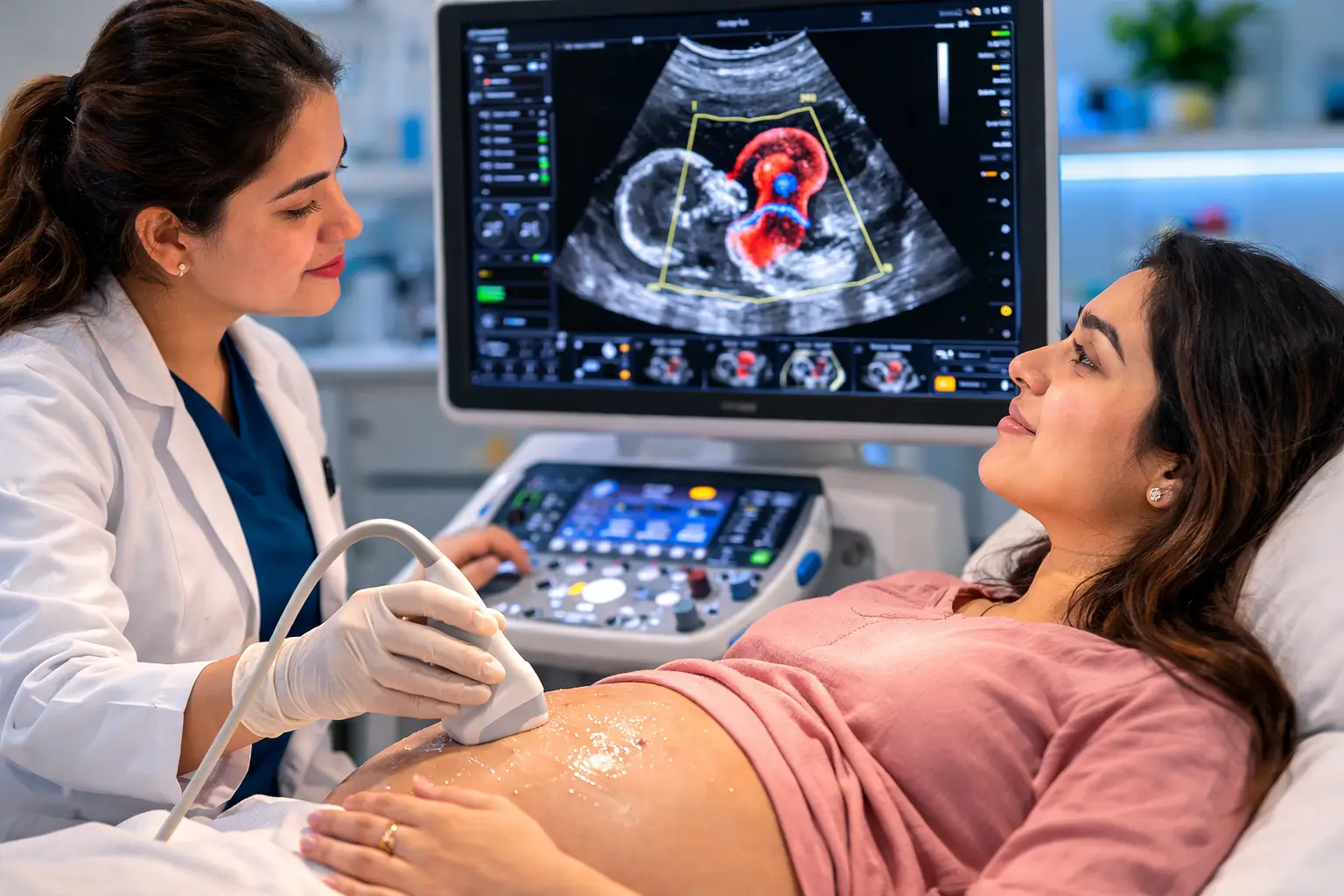
The Scan Itself
You lie on the examination table. Gel is applied to the abdomen. The sonographer uses a handheld transducer probe to obtain systematic views of the fetal heart. The scan typically takes 30–60 minutes. Depending on fetal position, the baby may need to move for optimal views — a short walk or glucose drink may be offered.
Views Obtained
Standard views include: 4-chamber view, 5-chamber/outflow view, 3-vessel and trachea view, aortic arch view, ductal arch view, bicaval view, short-axis views. Doppler is applied to assess blood flow velocity across valves and vessels.
M-Mode & Doppler Assessment
M-mode measures heart wall thickness and chamber sizes. Pulsed wave and colour Doppler assess valve regurgitation, septal defects, and flow patterns in great vessels.
Congenital Heart Defects Detected by Fetal Echo
- Ventricular Septal Defect (VSD) — hole between the lower chambers
- Atrial Septal Defect (ASD) — hole between the upper chambers
- Tetralogy of Fallot — combination of 4 heart defects
- Transposition of the Great Arteries (TGA)
- Hypoplastic Left Heart Syndrome (HLHS)
- Atrioventricular Septal Defect (AVSD) — common in Down syndrome
- Coarctation of the Aorta
- Ebstein’s Anomaly
- Complete Heart Block — fetal arrhythmia
- Cardiac tumours (rhabdomyoma)
What Do Fetal Echo Results Mean?
Normal Fetal Echo
All four chambers are of normal size and proportion. Valves open and close normally. No holes in the heart walls. Blood flow patterns are normal. Great arteries arise correctly. This is reassuring but does not guarantee the absence of all heart conditions.
Abnormal Fetal Echo
An abnormality does not always mean a serious problem. Many congenital heart defects (VSD, ASD) close spontaneously before birth or can be repaired after birth with excellent outcomes. Your fetal medicine specialist will explain the specific finding, its severity, and what to expect.
Fetal Echo Cost in Delhi 2026
At Edge Imaging, Fetal Echocardiography in Delhi is priced at ₹2,500–₹5,000 depending on the level of detail and Doppler assessment required. This is significantly more affordable than large private hospital rates of ₹8,000–₹15,000 for the same scan.
Frequently Asked Questions for Fetal Echocardiography in Delhi
Q1. When should I get a fetal echo?
Fetal echocardiography is ideally performed between 18–24 weeks. An early echo at 16–18 weeks is possible in very high-risk cases (e.g., previous child with CHD, maternal lupus) — though a repeat scan at 22–24 weeks may be needed for complete assessment.
Q2. Is fetal echo safe?
Yes — fetal echocardiography uses standard diagnostic ultrasound. No radiation is involved. Ultrasound has been used in obstetric imaging for 50+ years with an excellent safety record.
Q3. Does a normal fetal echo mean my baby definitely has no heart problem?
A normal fetal echo performed by an experienced operator provides very high reassurance. However, very small defects or those that develop later in pregnancy may not be detectable. Follow-up after birth is arranged for high-risk pregnancies.
Q4. What happens if fetal echo shows a heart problem?
You will be counselled by the fetal medicine specialist and referred to a paediatric cardiologist for further assessment and delivery planning. Many CHDs are manageable or correctable after birth. Pre-diagnosis allows delivery planning at a centre with neonatal cardiac surgery capability.
Q5. Is fetal echo covered by insurance?
Most insurance policies cover fetal echocardiography when ordered by a doctor for a specific indication. CGHS covers it at empanelled centres.
Q6. How long does fetal echo take?
A comprehensive Fetal Echocardiography in Delhi takes 30–60 minutes. In some cases (unfavourable fetal position), it may take longer or require you to return for a second attempt.
Q7. Can fetal echo detect Down syndrome?
Fetal echo detects the heart defect (AVSD) associated with Down syndrome, but it does not diagnose Down syndrome itself — that requires amniocentesis or NIPT for chromosomal analysis.
Q8. Where is the best fetal echo centre in West Delhi?
Edge Imaging & Diagnostics offers expert Fetal Echocardiography in Delhi at our Tagore Garden, Paschim Vihar, and Moti Nagar centres. Our high-resolution ultrasound systems and experienced radiologists deliver comprehensive cardiac assessment for your baby’s health.
Book Fetal Echocardiography at Edge Imaging Delhi
Your baby’s heart health matters. Edge Imaging’s expert Fetal Echocardiography in Delhi team provides detailed cardiac assessment in a comfortable, caring environment — giving you the answers and reassurance you need.
📍 Locations: Tagore Garden | Paschim Vihar | Moti Nagar | New Multan Nagar
✅ NABH Accredited | CGHS & DGHS Empanelled | Same-Day Reporting
📞 Call or WhatsApp to book your appointment today.
For more information, explore our related guide: Fetal Echo Test in Delhi
For more information on this procedure, refer to the World Health Organization (WHO) guidelines on medical procedures.
Medical Disclaimer: This article is for educational purposes only. Always consult a qualified medical professional for diagnosis and treatment advice.