
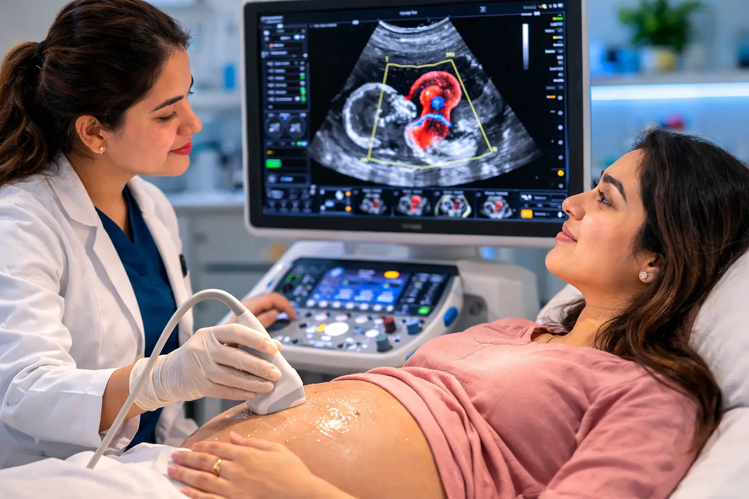
2D Echocardiography — commonly called a ‘2D echo’ or just ‘echo’ — is the most important and widely used test for evaluating the structure and function of the heart. Using harmless ultrasound waves, 2D echo provides real-time moving images of the heart that reveal its pumping function, valve health, wall motion, and chamber sizes. At Edge Imaging & Diagnostics in Delhi, we provide comprehensive 2D echo reports with same-day results.
Table of Contents
What Is 2D Echocardiography?
2D echocardiography is an ultrasound test that creates two-dimensional (flat, cross-sectional) images of the heart in real-time. The ‘2D’ refers to the two-dimensional plane of the image. By moving the transducer probe across the chest in standard positions, cardiologists and radiologists obtain multiple views of the heart — assessing every chamber, wall, and valve in detail.
What Does 2D Echo Show? for 2D Echocardiography in Delhi
Ejection Fraction (EF)
The most important measurement — the percentage of blood pumped out with each heartbeat. Normal: 55–70%. EF <40% indicates heart failure. EF 40–55% indicates mild impairment.
Wall Motion
Each segment of the heart wall is assessed for normal movement during contraction. Areas of reduced (hypokinesia), absent (akinesia), or paradoxical (dyskinesia) wall motion indicate areas damaged by heart attack (infarction).
Valve Assessment
All four valves (mitral, tricuspid, aortic, pulmonary) are assessed for stenosis (narrowing), regurgitation (leakage), and structural abnormalities. Doppler measurement of flow velocities quantifies the severity.
Chamber Sizes
Left ventricle, right ventricle, left atrium, and right atrium dimensions are measured. Enlargement of chambers indicates cardiac remodelling from chronic disease.
Pericardium
Fluid around the heart (pericardial effusion) is detected and quantified. Used to guide pericardiocentesis (drainage) if needed.
Aortic Root and Ascending Aorta
Aortic root dilation (aneurysm) is detected and measured — critical in Marfan syndrome and hypertension.
Left Ventricular Hypertrophy (LVH)
Thickening of the heart muscle from chronic hypertension — assessed by measuring septal and posterior wall thickness.
When Is 2D Echo Recommended? for 2D Echocardiography in Delhi
- Symptoms of heart disease: chest pain, breathlessness, palpitations, ankle swelling
- Newly diagnosed hypertension (to assess LVH and diastolic function)
- After a heart attack (myocardial infarction) — to assess extent of damage
- Known or suspected heart failure
- Evaluation of heart murmurs
- Pre-operative cardiac assessment
- Monitoring of known valve disease
- Suspected pericardial effusion
- Screening in athletes or persons with family history of heart disease
- Follow-up during chemotherapy (cardiotoxic drugs)
2D Echo Report: Key Values Explained
Ejection Fraction (EF)
Normal: 55–70%. Mildly reduced: 45–54%. Moderately reduced: 30–44%. Severely reduced: <30%.
LV Internal Diameter (diastole/systole)
Normal LVIDD: 3.5–5.5 cm. Enlarged: >5.5 cm (dilated cardiomyopathy).
Interventricular Septum (IVS)
Normal: 0.6–1.1 cm. Thickened: >1.2 cm (hypertensive heart disease, HCM).
Mitral Valve
E/A ratio >1 = normal diastolic function. E/A <1 = diastolic dysfunction. Mitral E/A, E' wave, and E/E' ratio assess diastolic dysfunction grade.
LVEF by Simpson’s Method
Most accurate ejection fraction measurement. Uses apical 4-chamber and 2-chamber views with manual tracing. Standard in all modern echo reports.
Types of Echocardiography at Edge Imaging
- 2D Echo: Standard real-time cross-sectional imaging
- M-Mode Echo: One-dimensional time-motion study for precise measurements
- Colour Doppler: Visualises blood flow direction in the heart
- Pulsed Wave Doppler: Measures velocity at specific points
- Continuous Wave Doppler: Measures high-velocity flows across valves
- Tissue Doppler Imaging (TDI): Assesses myocardial velocity for diastolic function
2D Echo Cost in Delhi 2026
At Edge Imaging, 2D echocardiography is priced at ₹1,200–₹2,500 depending on the level of assessment (standard echo vs full Doppler study). CGHS/DGHS rates available. Same-day reporting. Compare: large private hospitals charge ₹4,000–₹8,000 for the identical test.
How to Prepare for 2D Echo
- No fasting required for standard transthoracic 2D echo
- Avoid heavy meals immediately before the scan (minor personal comfort measure only)
- Wear comfortable, loose two-piece clothing
- Remove necklaces and chest jewellery
- Continue all cardiac medications as normal
- Bring all previous ECG, echo reports, and prescription to appointment
Frequently Asked Questions
Q1. How long does a 2D echo take?
A comprehensive 2D echo with Doppler typically takes 20–30 minutes for the scan. The report is ready within a few hours of the scan at Edge Imaging.
Q2. Is 2D echo the same as an ECG?
No. ECG (electrocardiogram) records the electrical activity of the heart — showing rhythm and conduction patterns. 2D echo shows the structural and functional anatomy — how the heart looks and pumps. Both are complementary tests.
Q3. What is a normal ejection fraction on 2D echo?
A normal ejection fraction (EF) is 55–70%. An EF of 50–55% is considered borderline. Below 50% requires further evaluation for the cause of impairment. Below 35% indicates significant heart failure.
Q4. Can 2D echo detect blocked arteries?
2D Echocardiography in Delhi does not directly visualise coronary arteries. However, it can detect indirect signs of coronary artery disease (CAD) — areas of reduced wall motion resulting from previous heart attacks. A TMT test or stress echo is better for detecting active CAD.
Q5. Do I need 2D echo before surgery?
Pre-operative 2D echo is recommended for patients with known heart disease, symptoms suggestive of cardiac dysfunction, or those undergoing major surgery. Your anaesthesiologist or surgeon will specify if it is required for your procedure.
Q6. How often should I repeat 2D echo?
For known heart disease: as advised by your cardiologist (usually every 6–12 months). For hypertension monitoring: every 1–2 years. For pre-operative assessment: single test if no prior cardiac history.
Q7. Is 2D echo safe?
Completely safe. 2D Echocardiography in Delhi uses non-ionising ultrasound waves — no radiation, no needles. It is safe for pregnant women, children, and the elderly.
Q8. What is the difference between 2D echo and fetal echo?
2D echo examines an adult’s or child’s heart after birth. Fetal echocardiography specifically examines an unborn baby’s heart during pregnancy using specialised views and protocols. Both are ultrasound-based but optimised for their specific use.
Book 2D Echo at Edge Imaging Delhi — Same-Day Report
Heart health assessment should never be delayed. Edge Imaging’s 2D Echocardiography in Delhi service delivers comprehensive cardiac imaging with same-day reporting at West Delhi’s most affordable prices.
📍 Locations: Tagore Garden | Paschim Vihar | Moti Nagar | New Multan Nagar
✅ NABH Accredited | CGHS & DGHS Empanelled | Same-Day Reporting
📞 Call or WhatsApp to book your appointment today.
For more information, explore our related guide: Fetal Echo Test in Delhi
For more information on this procedure, refer to the World Health Organization (WHO) guidelines on medical procedures.
Medical Disclaimer: This article is for educational purposes only. Always consult a qualified medical professional for diagnosis and treatment advice.