
A fetal echo test in Delhi (foetal echocardiography) is a specialised ultrasound examination that provides a detailed assessment of the developing baby’s heart during pregnancy. If your obstetrician has referred you for a fetal echo test in Delhi, understanding what this important scan involves will help ease any anxiety and ensure you are well-prepared. At Edge Imaging & Diagnostics in Paschim Vihar, West Delhi, our expert sonologists perform fetal echo test in Delhi using high-resolution ultrasound technology to provide accurate assessment of your baby’s cardiac health.
Congenital heart disease (CHD) is the most common type of birth defect, affecting approximately 8 in 1,000 live births. A fetal echo test in Delhi allows early detection of structural heart abnormalities before birth, enabling planning for optimal delivery and immediate neonatal cardiac care when needed. Expectant mothers choosing Edge Imaging & Diagnostics for their fetal echo test in Delhi benefit from expert reporting and compassionate care throughout their pregnancy journey.
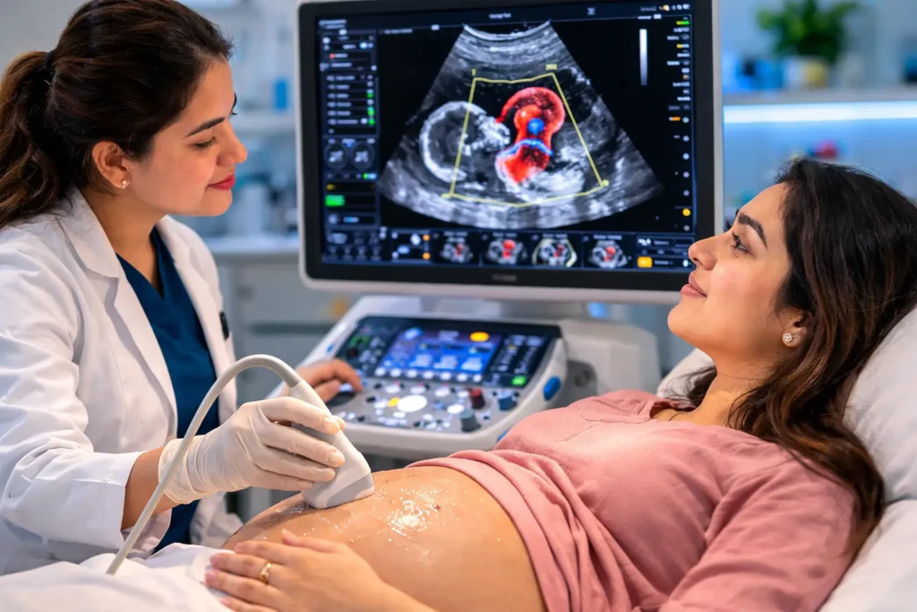
Table of Contents
What is a Fetal Echo Test?
A fetal echocardiography test (Fetal Echo) is a detailed ultrasound examination of the foetal heart performed during pregnancy to assess the structure, function, rhythm, and blood flow of the developing baby’s heart. It uses the same high-frequency sound wave technology as a standard obstetric ultrasound but focuses specifically on the heart with specialised cardiac imaging protocols.
The fetal echo test evaluates all four chambers of the heart, the valves, the great vessels (aorta and pulmonary artery), the connections between heart chambers, and the direction and velocity of blood flow using Doppler technology. This comprehensive cardiac evaluation allows detection of structural heart defects (congenital heart disease) that may need medical or surgical intervention after birth.
Fetal echocardiography is typically performed between 18–24 weeks of pregnancy — the optimal time window when the foetal heart is large enough for detailed imaging but early enough in pregnancy to allow counselling and delivery planning. In certain high-risk cases, an early fetal echo may be attempted at 12–14 weeks using a transvaginal approach.
Why is a Fetal Echo Test Done?
Fetal echocardiography is performed to detect congenital heart defects and assess foetal cardiac function. Key reasons it is ordered include:
- Abnormal four-chamber view on anomaly scan: When the routine fetal anomaly scan (Level 2 ultrasound) shows an abnormal or suspicious cardiac view requiring detailed evaluation.
- Family history of congenital heart disease: If a parent or previous sibling had a congenital heart defect, the risk of recurrence is increased.
- Maternal diabetes mellitus: Pre-gestational diabetes (Type 1 or Type 2) significantly increases the risk of foetal congenital heart disease.
- Maternal rubella or viral infection in first trimester: Certain infections during early pregnancy are associated with foetal cardiac defects.
- Maternal medications: Some drugs taken in pregnancy (lithium, phenytoin, warfarin) are associated with increased cardiac defect risk.
- Abnormal foetal karyotype: Chromosomal abnormalities like Down syndrome (Trisomy 21), Turner syndrome, and DiGeorge syndrome are associated with specific cardiac defects.
- Increased NT (Nuchal Translucency) on first trimester scan: Increased NT is associated with both chromosomal abnormalities and structural heart defects.
- Foetal arrhythmia detected on routine scan: Irregular foetal heart rate or rhythm requires detailed echocardiographic assessment.
- Maternal connective tissue disease: Systemic lupus erythematosus (SLE) with anti-Ro/La antibodies can cause complete heart block in the foetus.
- Twin pregnancy: Higher risk of cardiac abnormalities, particularly in monochorionic twins with twin-to-twin transfusion syndrome.
Who Needs a Fetal Echo Test?
A fetal echo test in Delhi is recommended for pregnant women who have:
- Pre-gestational diabetes mellitus (Type 1 or Type 2)
- A personal or family history of congenital heart disease
- A previous pregnancy affected by congenital heart disease
- Abnormal cardiac views on the routine anomaly scan
- Increased nuchal translucency (NT > 3.5 mm) on first trimester screening
- Confirmed foetal chromosomal abnormality (Down syndrome, Turner syndrome)
- Maternal rubella, CMV, or other relevant infection in the first trimester
- Maternal SLE with anti-Ro antibodies
- Teratogenic drug exposure in the first trimester
- Foetal hydrops (fluid accumulation in body cavities) detected on ultrasound
- Multiple gestation with monochorionic placentation
When is a Fetal Echo Test Advised?
Your obstetrician may specifically recommend a fetal echo scan in Delhi if:
- The routine level 2 anomaly scan showed a suspected cardiac abnormality
- The foetal heart rate is irregular or persistently slow (bradycardia) or fast (tachycardia) on routine check
- You have pre-existing diabetes and have not yet had a detailed foetal cardiac scan
- A close family member was born with a heart defect
- Your first trimester blood tests or NT scan showed increased Down syndrome risk
Preparation Before a Fetal Echo Test
Fetal echocardiography requires minimal preparation:
- No fasting required: You can eat and drink normally before the scan.
- Full bladder not required in most cases (unlike early pregnancy scans). A partially filled bladder may help visualise the foetal position.
- Timing: The ideal time for fetal echo is between 18–24 weeks of gestation. Early fetal echo at 12–14 weeks may be done transvaginally or transabdominally.
- Documents: Bring your antenatal record, previous ultrasound reports, and the referral from your obstetrician.
- Clothing: Wear comfortable, loose-fitting clothing that allows easy access to the abdomen.
- Companion: You are welcome to bring a partner or family member for support and to share in the experience.
Step-by-Step Fetal Echo Test Procedure
Here is a detailed walkthrough of the fetal echo test procedure at Edge Imaging & Diagnostics in Paschim Vihar, Delhi:
- Patient positioning: You lie comfortably on the examination table on your back. A pillow is placed under your knees for comfort.
- Gel application: Warm ultrasound gel is applied to your abdomen to enable good acoustic coupling between the probe and skin.
- Foetal position assessment: The sonologist first assesses foetal position, lie, and basic anatomy before focusing on the heart.
- Cardiac anatomy imaging: The sonologist systematically evaluates the four-chamber view, left and right ventricular outflow tracts, aortic and ductal arches, superior and inferior vena cava connections, and pulmonary venous return.
- Colour flow Doppler: Colour Doppler mapping is applied to assess the direction and velocity of blood flow across all four valves and through the great vessels — detecting abnormal flow patterns caused by stenosis, regurgitation, or septal defects.
- Pulsed wave Doppler: Quantitative flow velocities are measured across the atrioventricular valves (mitral and tricuspid) and semilunar valves (aortic and pulmonary) to assess valve function and cardiac output.
- Foetal heart rhythm assessment: The cardiac rhythm is assessed for rate, regularity, and atrioventricular synchrony — identifying any arrhythmia present.
- M-mode echocardiography: Used to measure cardiac dimensions and assess ventricular function (fractional shortening, cardiac contractility).
- Documentation: All key views and measurements are recorded, documented, and archived.
- Reporting: The sonologist prepares a comprehensive fetal echo report describing all cardiac structures, function, and any abnormalities identified.
Total examination time is typically 30–45 minutes. In some cases, foetal position may require repositioning or rescanning after a short walk to obtain optimal cardiac views.
What Happens After a Fetal Echo Test?
- You can resume all normal activities immediately after the scan.
- The fetal echo report is typically ready the same day at Edge Imaging & Diagnostics.
- A normal fetal echo provides reassurance that the baby’s heart structure and function appear normal for gestational age.
- If an abnormality is detected, your obstetrician will refer you to a paediatric cardiologist for further counselling, additional investigations, and birth planning at a tertiary centre with neonatal cardiac surgery capability.
- Fetal echo may be repeated at 28–30 weeks if an earlier scan was technically limited or if a monitored condition requires surveillance.
Safety of Fetal Echo Test
Fetal echocardiography is completely safe for both the mother and the developing baby. It uses only ultrasound — no radiation, no needles, no contrast agents. Ultrasound has been used safely in obstetric practice for over 50 years with no adverse effects demonstrated on foetal development. The procedure is non-invasive and causes no discomfort beyond the pressure of the ultrasound probe on the abdomen.
Benefits of Fetal Echo Test
- Early detection of congenital heart disease before birth — enabling planned delivery at an appropriate tertiary centre
- Allows time for parental counselling and psychological preparation
- Guides neonatal management immediately after birth — the cardiac team is prepared in advance
- Identifies foetal cardiac arrhythmias treatable with maternal medication during pregnancy
- Detects conditions (like complete heart block in SLE mothers) allowing preventive treatment
- Normal result provides enormous reassurance to high-risk parents
- Completely safe — no radiation, no risk to baby
Fetal Echo Test Accuracy
Fetal echocardiography performed between 18–24 weeks by an experienced sonologist achieves a detection sensitivity of 70–85% for major structural congenital heart defects. The detection rate for severe defects such as hypoplastic left heart syndrome, transposition of the great arteries, and tetralogy of Fallot is above 90% in specialised centres. Accuracy depends significantly on operator expertise, image quality, foetal position, and maternal body habitus. At Edge Imaging & Diagnostics in Paschim Vihar, Delhi, our sonologists are trained in foetal cardiac imaging protocols and perform detailed systematic fetal echo examinations to maximise diagnostic accuracy.
Cost of Fetal Echo Test in Delhi
| Fetal Echo Type | Approximate Cost in Delhi |
|---|---|
| Standard Fetal Echocardiography (18–24 weeks) | ₹2,500 – ₹6,000 |
| Early Fetal Echo (12–14 weeks, transvaginal) | ₹3,000 – ₹7,000 |
| Detailed Fetal Echo with Colour Doppler | ₹4,000 – ₹8,000 |
At Edge Imaging & Diagnostics in West Delhi, fetal echo scans are offered at competitive prices with complete transparency. We accept all major health insurance and TPA cashless facilities. Patients from Paschim Vihar, Punjabi Bagh, Rajouri Garden, and across Delhi NCR are welcome.
Why Choose Edge Imaging & Diagnostics for Fetal Echo in Delhi?
- High-resolution ultrasound equipment with advanced foetal cardiac imaging capability
- Experienced sonologists trained in systematic fetal echocardiography protocols
- Comprehensive assessment including 2D, colour Doppler, pulsed wave Doppler, and M-mode imaging
- Detailed structured reports prepared same day
- Warm, welcoming environment for expecting mothers — companions welcome
- Conveniently located in Paschim Vihar — accessible from Punjabi Bagh, Rajouri Garden, and all of Delhi NCR
- Insurance and TPA cashless accepted
- Transparent pricing with no hidden charges
Frequently Asked Questions About Fetal Echo Test
1. Is the fetal echo test safe for my baby?
Yes. Fetal echocardiography is completely safe for both mother and baby. It uses only ultrasound waves — no radiation or contrast agents. Ultrasound has been used safely in obstetric imaging for over 50 years without any demonstrated adverse effects on foetal development.
2. When is the best time for a fetal echo test?
The ideal time for fetal echocardiography is between 18–24 weeks of gestation. At this stage, the foetal heart is large enough for detailed evaluation. Early fetal echo at 12–14 weeks is possible but has lower sensitivity and is reserved for specific high-risk indications.
3. How long does a fetal echo test take?
A fetal echo scan typically takes 30–45 minutes. If foetal position is suboptimal, a short walk may help reposition the baby, and the scan may take slightly longer to obtain all required views.
4. What heart defects can fetal echo detect?
Fetal echo can detect ventricular and atrial septal defects (VSD, ASD), hypoplastic left heart syndrome, transposition of great arteries, tetralogy of Fallot, pulmonary and aortic stenosis, coarctation of the aorta, complete atrioventricular canal defects, Ebstein anomaly, and cardiac arrhythmias.
5. Does a normal fetal echo guarantee a healthy heart?
A normal fetal echo provides very strong reassurance but does not provide a 100% guarantee. Small VSDs, some cardiac rhythm issues, and minor valve abnormalities can occasionally be missed. Postnatal examination by a paediatrician remains essential for all newborns.
6. Is fetal echo different from a routine anomaly scan?
Yes. A routine anomaly scan (Level 2 or morphology scan) provides an overview of foetal anatomy including a basic four-chamber cardiac view. Fetal echocardiography is a dedicated, much more detailed examination of the heart using multiple specialised views and Doppler techniques that routine anomaly scans do not include.
7. Do I need to fast before a fetal echo test?
No fasting is required for a fetal echo scan. You can eat and drink normally. Wearing loose, comfortable clothing that allows easy access to your abdomen is recommended.
8. What happens if a heart defect is found on fetal echo?
If a cardiac abnormality is detected, you will be referred to a paediatric cardiologist for detailed counselling. Most congenital heart defects are surgically correctable. Knowing about the defect before birth allows delivery to be planned at a centre with neonatal cardiac surgery capability, dramatically improving outcomes.
9. I have diabetes in pregnancy — do I need a fetal echo?
Yes. Pre-gestational diabetes (Type 1 or Type 2 diagnosed before pregnancy) significantly increases the risk of congenital heart disease in the baby. Current guidelines recommend fetal echocardiography for all women with pre-gestational diabetes. Gestational diabetes (developing during pregnancy) carries a lower risk but may also warrant a fetal echo.
10. How do I book a fetal echo test in Delhi?
Book your fetal echocardiography at Edge Imaging & Diagnostics in Paschim Vihar, West Delhi by calling our centre or visiting edgeimaging.in. Bring your antenatal records, obstetrician’s referral, and previous ultrasound scan reports.
11. Can fetal echo detect Down syndrome?
Fetal echo cannot diagnose Down syndrome (which requires chromosomal testing), but it can detect the cardiac defects that are commonly associated with Down syndrome — particularly atrioventricular septal defects (AVSDs) and VSDs. A normal fetal echo does not rule out Down syndrome.
12. Is fetal echo covered by health insurance?
Fetal echocardiography is covered by most maternity health insurance policies in India when performed for a medically indicated high-risk pregnancy. Edge Imaging & Diagnostics in Paschim Vihar accepts all major insurance and TPA cashless facilities.
Best Diagnostic Centre for Fetal Echo Test in Delhi
When searching for the best centre for a fetal echo test in Delhi, expectant parents need a facility with advanced fetal echocardiography equipment and experienced sonologists. Edge Imaging & Diagnostics in Paschim Vihar, West Delhi, is the premier choice for fetal echo test in Delhi, serving patients from Punjabi Bagh, Rajouri Garden, and across Delhi NCR. Our experienced team performs every fetal echo test in Delhi with the utmost care, providing detailed reports on fetal cardiac anatomy and function. Book your fetal echo test in Delhi at Edge Imaging & Diagnostics for expert foetal cardiac assessment and peace of mind during pregnancy.
Medical References: For evidence-based guidelines on foetal echocardiography, refer to the American Institute of Ultrasound in Medicine (AIUM) — Foetal Echocardiography Practice Guidelines and the International Society of Ultrasound in Obstetrics & Gynecology (ISUOG) Fetal Echo Guidelines.
Conclusion
A fetal echo test in Delhi is an invaluable diagnostic examination that provides comprehensive assessment of your baby’s heart health before birth. For high-risk pregnancies, early detection of congenital heart disease allows optimal birth planning, immediate neonatal cardiac care, and in many cases, lifesaving surgical intervention after delivery.
At Edge Imaging & Diagnostics in Paschim Vihar, West Delhi, our experienced sonologists perform detailed fetal echocardiography using state-of-the-art equipment, with same-day structured reporting and compassionate care for every expecting mother. We serve patients from Punjabi Bagh, Rajouri Garden, Moti Nagar, and throughout Delhi NCR.
Book your fetal echo test in Delhi today at Edge Imaging & Diagnostics — because every baby deserves a strong start.
Also read: Ultrasound Scan in Delhi | Open MRI Scan in Delhi | Amniocentesis Test in Delhi