
Ultrasound-Guided FNAC in Delhi — When a lump or mass is too small, too deep, or cannot be clearly felt by touch alone, ultrasound-guided FNAC (USG-FNAC) is the gold standard diagnostic approach. By using real-time ultrasound imaging to guide the needle to the exact lesion, the radiologist ensures maximum accuracy while avoiding surrounding vital structures.
Table of Contents
What Is Ultrasound-Guided FNAC?
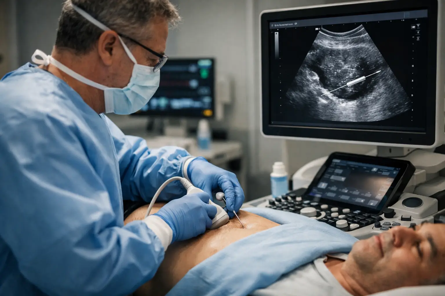
Ultrasound-guided FNAC (also called USG-guided FNAC or image-guided FNAC) is a procedure in which a fine needle is inserted into a lesion under continuous real-time ultrasound visualisation. The radiologist watches the needle on the ultrasound screen at every moment, confirming that the tip is inside the target lesion before aspirating cells. This eliminates guesswork and dramatically improves the yield of diagnostic material.
How Is USG-Guided FNAC Different from Blind FNAC? for Ultrasound-Guided FNAC in Delhi
Blind (Palpation-Guided) FNAC
The doctor inserts the needle based on feeling the lump. Suitable for surface lumps that are easily felt. Less accurate for small (<1 cm), deep, or partially cystic lesions. Risk of missing the target or sampling the non-representative part of the lesion.
Ultrasound-Guided FNAC
The needle is inserted under real-time ultrasound visualisation. The radiologist sees the needle tip inside the lesion on screen. Suitable for all lesions — including those too small or deep to feel. Achieves 95%+ accuracy and significantly reduces non-diagnostic samples.
Which Lesions Require USG-Guided FNAC? for Ultrasound-Guided FNAC in Delhi
- Thyroid nodules (especially those <1.5 cm or partially cystic)
- Breast lumps not clearly palpable on physical examination
- Liver masses, hepatic lesions, or liver abscesses
- Neck lymph nodes that are deep or near important vessels
- Axillary lymph nodes in breast cancer staging
- Renal (kidney) masses
- Pancreatic lesions accessible via endoscopic ultrasound
- Ovarian or pelvic masses
- Soft tissue tumours with mixed solid-cystic components
USG-Guided FNAC Procedure at Edge Imaging
Step 1: Pre-Procedure Ultrasound
The radiologist performs a diagnostic ultrasound to locate, measure, and characterise the target lesion. The skin entry point, needle angle, and depth are planned. Vital structures (blood vessels, nerves) are mapped and avoided.
Step 2: Skin Preparation & Anaesthesia
The skin is cleaned with antiseptic. For surface-level lesions, no anaesthesia is needed. For deep lesions or anxious patients, 1–2 ml of local lignocaine is injected subcutaneously to numb the entry point.
Step 3: Needle Insertion Under Real-Time Guidance
The fine needle is inserted through the skin while the radiologist watches the ultrasound screen. The needle tip appears as a bright echogenic dot on the screen, allowing the radiologist to steer it precisely into the lesion.
Step 4: Aspiration
Once the needle tip is confirmed inside the target lesion, negative pressure is applied via the syringe to aspirate cells. The needle is gently moved back and forth within the lesion to collect cells from different areas.
Step 5: Sample Preparation
The aspirated material is expelled onto glass slides, smeared, fixed in alcohol or air-dried, and sent to the pathology laboratory. The procedure is repeated 2–3 times (passes) to ensure adequate cellularity.
Step 6: Report & Consultation
The cytology report is ready within 24–48 hours. At Edge Imaging, a free doctor consultation is provided after your result to explain the findings and discuss next steps.
Benefits of USG-Guided FNAC at Edge Imaging
- Real-time visualisation ensures needle reaches the exact target
- 95%+ diagnostic accuracy vs 70–80% for palpation-guided FNAC
- Significantly reduced rate of non-diagnostic (inadequate) samples
- Safe avoidance of blood vessels and adjacent structures
- Can sample specific areas within a complex lesion (solid vs cystic)
- Faster procedure with less patient discomfort
- Immediate confirmation of adequate sample before patient leaves
- Available at all Edge Imaging West Delhi centres
USG-Guided FNAC Cost in Delhi 2026
At Edge Imaging, ultrasound-guided FNAC is priced between ₹1,500 and ₹3,500 depending on the organ/site. This includes the ultrasound guidance, procedure, and pathology report. CGHS/DGHS rates are available for eligible patients.
Technology Used at Edge Imaging for USG-FNAC
We use high-resolution Doppler ultrasound systems capable of producing crystal-clear images of lesions as small as 5mm. Colour Doppler is used to map blood vessels before needle insertion, ensuring the safest possible approach. Our systems are regularly calibrated and maintained to the highest imaging standards.
Frequently Asked Questions
Q1. Is USG-guided FNAC painful?
Most patients experience only mild discomfort — similar to a blood test. For deep lesions, local anaesthetic is applied first. The entire procedure including preparation takes approximately 30–45 minutes.
Q2. How accurate is ultrasound-guided FNAC?
In experienced hands, USG-guided FNAC achieves 93–97% sensitivity for malignancy. This is significantly higher than palpation-guided FNAC (70–85%) because image guidance ensures the needle samples the most representative area of the lesion.
Q3. Can USG-guided FNAC diagnose all types of cancer?
USG-guided FNAC can identify most cancer types at the cytological level. It may not always provide enough tissue for full histological characterisation (e.g., lymphoma subtyping or receptor testing in breast cancer), in which case core needle biopsy may be additionally recommended.
Q4. Do I need to fast before USG-guided FNAC?
For thyroid, neck, or breast FNAC, no fasting is needed. For abdominal lesions (liver, kidney, pancreas), 4–6 hours of fasting is advised. Your appointment confirmation will specify requirements.
Q5. How many passes are needed in USG-guided FNAC?
Typically 2–4 passes (needle insertions) are performed to ensure adequate cellularity. The radiologist assesses the material at each pass and stops when sufficient cells have been collected.
Q6. When will I get my USG-FNAC report?
At Edge Imaging, most Ultrasound-Guided FNAC in Delhi reports are available within 24 hours. Same-day preliminary results are available in urgent cases.
Q7. Is USG-guided FNAC available in West Delhi?
Yes, all Edge Imaging centres in West Delhi — Tagore Garden, Paschim Vihar, and Moti Nagar — offer Ultrasound-Guided FNAC in Delhi by experienced radiologists.
Q8. What is ROSE in FNAC?
ROSE (Rapid On-Site Evaluation) is a technique where a cytotechnician or pathologist examines the slide immediately after aspiration to confirm adequate sampling before the patient leaves. This reduces the risk of needing a repeat procedure. Ask Edge Imaging about ROSE availability for your procedure.
Book USG-Guided FNAC at Edge Imaging West Delhi
Get the most accurate Ultrasound-Guided FNAC in Delhi result with real-time guidance at Edge Imaging & Diagnostics. Our experienced interventional radiologists and same-day reporting ensure you get answers quickly and confidently.
📍 Locations: Tagore Garden | Paschim Vihar | Moti Nagar | New Multan Nagar
✅ NABH Accredited | CGHS & DGHS Empanelled | Same-Day Reporting
📞 Call or WhatsApp to book your appointment today.
For more information, explore our related guide: FNAC Test in Delhi
For more information on this procedure, refer to the World Health Organization (WHO) guidelines on medical procedures.
Medical Disclaimer: This article is for educational purposes only. Always consult a qualified medical professional for diagnosis and treatment advice.